38-year-old patient with progressive dyspnea and cough
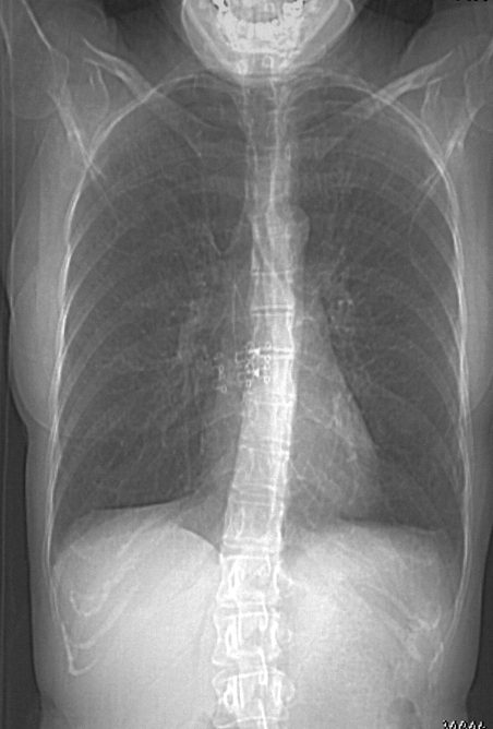
CXR (scout for CT) shows hyperinflated lungs with increased lung volumes
38-year-old patient with progressive dyspnea and cough
CXR (scout for CT) shows hyperinflated lungs with increased lung volumes
Ashley Davidoff MD
CT shows bilateral and extensive thin-walled cysts surrounded by very little normal lung parenchyma.
The cysts are round and thin-walled except for air filled large irregular pocket in the right apex (image 27628/29) .
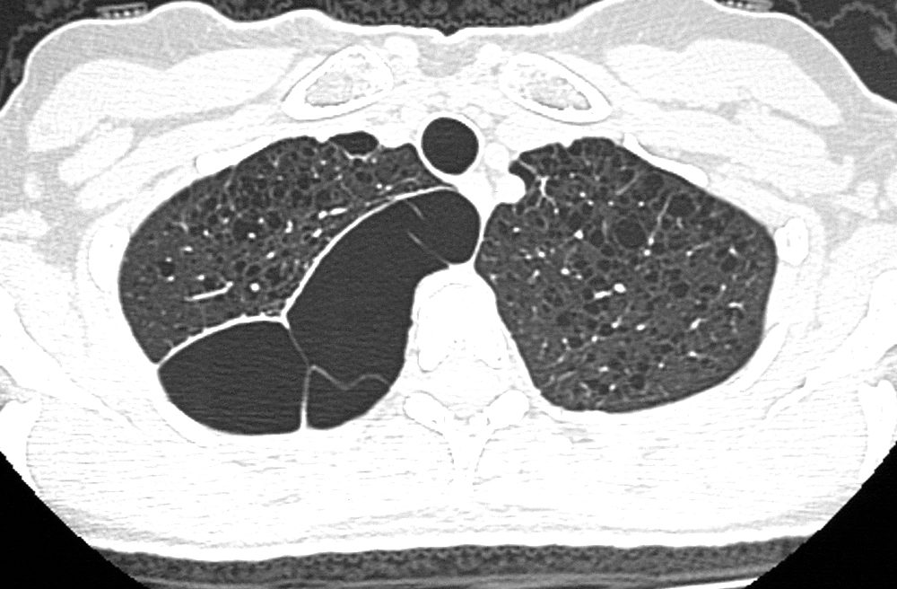
The cysts are round and thin-walled except for air filled large irregular pocket in the right apex (image 27628/29) .
Ashley Davidoff MD
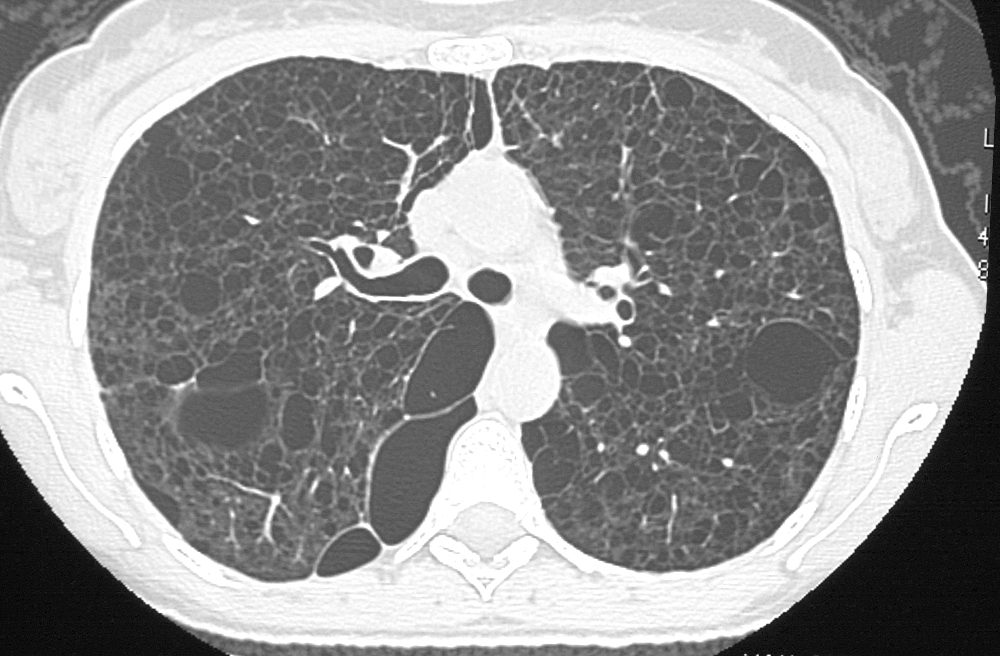
The cysts are round and thin-walled except for air filled large irregular pocket in the right apex (image 27628/29) .
Ashley Davidoff MD
Some of the cysts do not have walls at all.
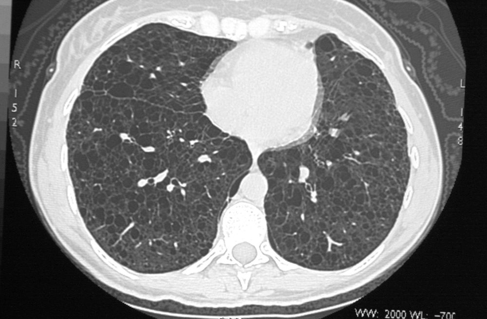
Some of the cysts do not have walls at all and others have an irregular configuration
Ashley Davidoff MD
In the abdomen multiple low density lymphangioleiomyomas are present that are due to lymphatic obstruction.
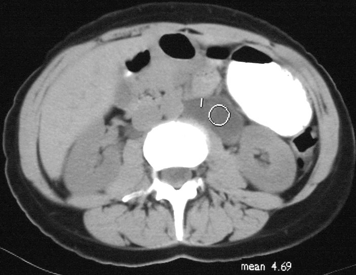
In the abdomen multiple low density lymphangioleiomyomas are present that are due to lymphatic obstruction.
Ashley Davidoff MD
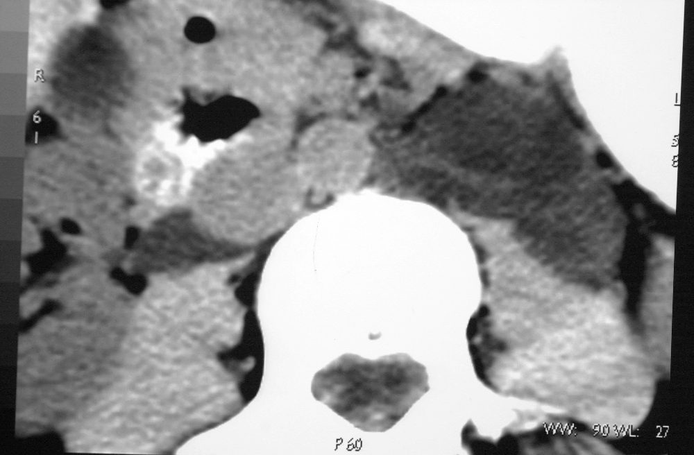
In the abdomen multiple low density lymphangioleiomyomas are present that are due to lymphatic obstruction.
Ashley Davidoff MD
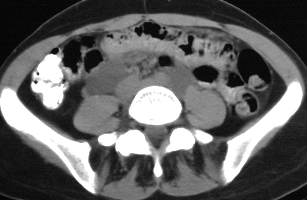
In the abdomen multiple low density lymphangioleiomyomas are present that are due to lymphatic obstruction.
Ashley Davidoff MD
