211Lu Adenocarcinoma Huge Necrotic Mass

53 year old male  history of stage IIIB adeno carcinoma of the left upper lobe with prevascular and AP window lymph node involvement based on PET/CT with presumed lymphangitic spread surrounding the large mass. Treatment has included both radiation and chemotherapy
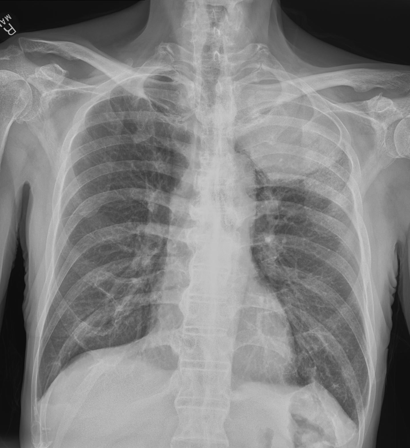
53 year old male with history of smoking presents with a cough CXR shows a large mass in the left apex
Diagnosis – adenocarcinoma of the lung with extensive necrosis of the tumor
Ashley Davidoff MD The CommonVein.net
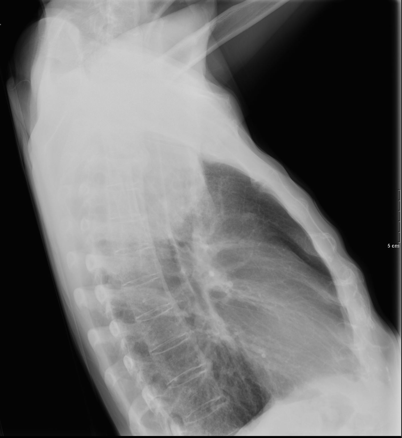
53 year old male with history of smoking presents with a cough CXR shows a large mass in the left apex
Diagnosis – adenocarcinoma of the lung with extensive necrosis of the tumor
Ashley Davidoff MD The CommonVein.net
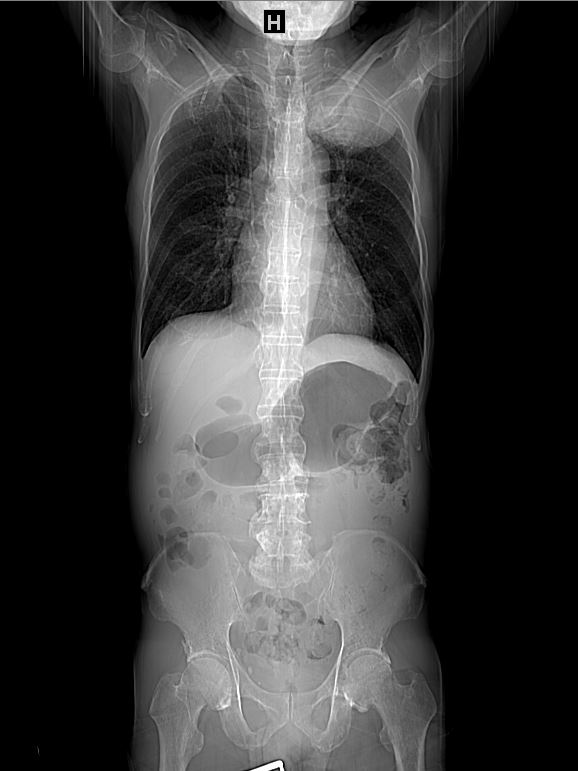
53 year old male with history of smoking presents with a cough Scout for the CT scan shows a large mass  shows a large mass in the left apex
Diagnosis – adenocarcinoma of the lung with extensive necrosis of the tumor
Ashley Davidoff MD The CommonVein.net
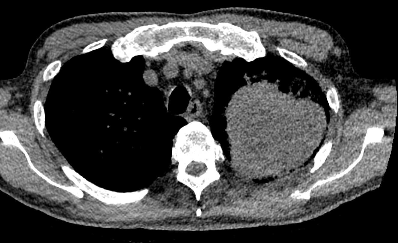
53 year old male with history of smoking presents with a cough Axial CT scan with soft tissue windows shows a 8.5 cm mass in the left upper lobe
Diagnosis – adenocarcinoma of the lung with extensive necrosis of the tumor
Ashley Davidoff MD The CommonVein.net
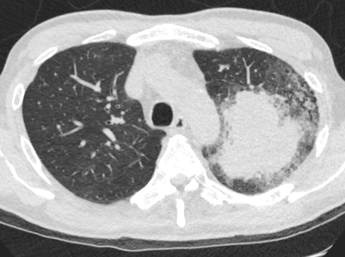
53 year old male with history of smoking presents with a cough Axial CT scan with lung windows windows shows the mass with  surrounding ground glass changes with thickening of interlobular septa and thickening of adjacent airways reminiscent of lymphangitis
Diagnosis – adenocarcinoma of the lung with extensive necrosis of the tumor
Ashley Davidoff MD The CommonVein.net
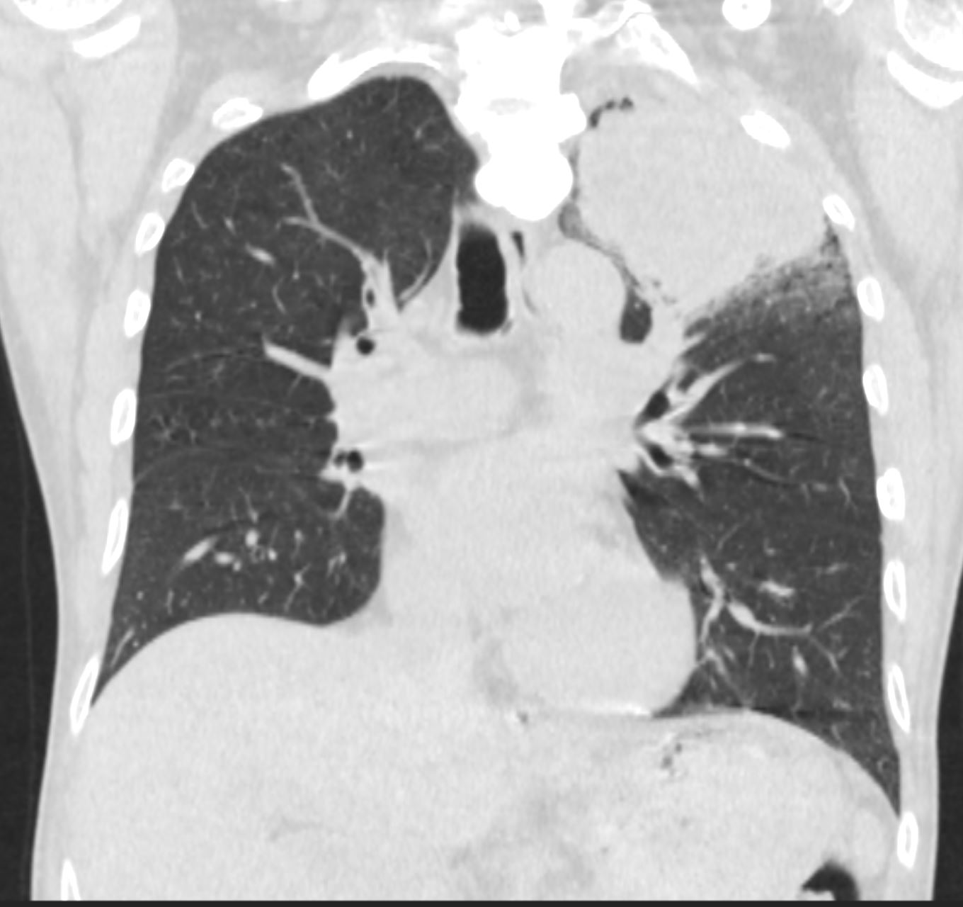
53 year old male with history of smoking presents with a cough Coronal reconstruction of the CT scan with lung windows windows shows the mass with surrounding ground glass changes with thickening of interlobular septa and thickening of adjacent airways reminiscent of lymphangitis
Diagnosis – adenocarcinoma of the lung with extensive necrosis of the tumor
Ashley Davidoff MD The CommonVein.net
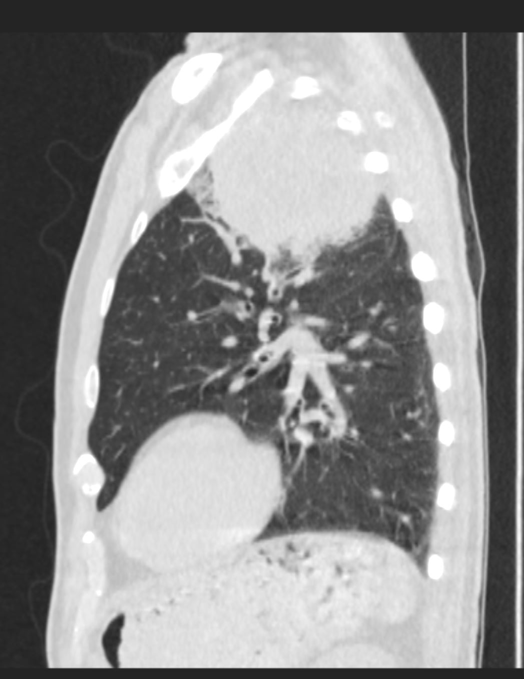
53 year old male with history of smoking presents with a cough Sagittal reconstruction of the CT scan with lung windows windows shows the mass with surrounding ground glass changes with thickening of interlobular septa and thickening of adjacent airways reminiscent of lymphangitis
Diagnosis – adenocarcinoma of the lung with extensive necrosis of the tumor
Ashley Davidoff MD The CommonVein.net
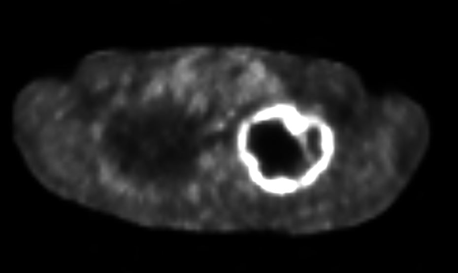
 

53 year old male with history of smoking presents with a cough PET CT shows a large 8.9 cm left upper lobe apical lung mass which  is peripherally intensely hypermetabolic, centrally photopenic there is suspicious for lung malignancy with central necrosis  The tumor circumferentially abuts the pleura and abuts the anterior and posterior upper ribs and upper T-spine without low-dose CT evidence of erosion.
Diagnosis – adenocarcinoma of the lung with extensive necrosis of the tumor
Ashley Davidoff MD The CommonVein.net
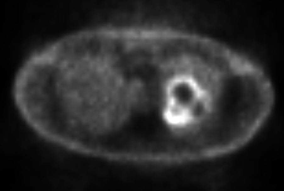
53 year old male with history of smoking presents with a cough PET CT shows a large 8.9 cm left upper lobe apical lung mass which  is peripherally intensely hypermetabolic, centrally photopenic there is suspicious for lung malignancy with central necrosis  The tumor circumferentially shows mild tracer uptake and becomes suspicious for lymphangitis carcinomatosa
Diagnosis – adenocarcinoma of the lung with extensive necrosis of the tumor
Ashley Davidoff MD The CommonVein.net