6 years prior
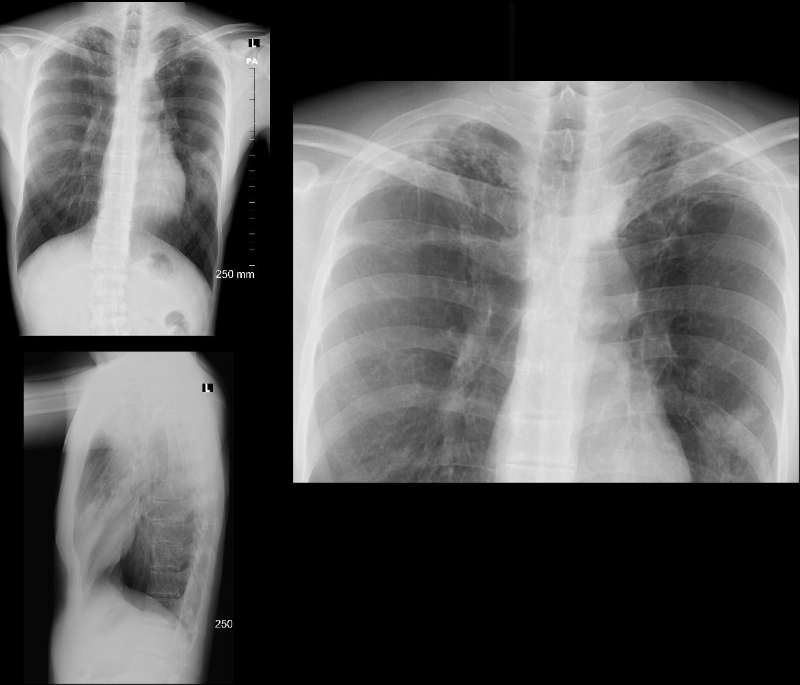
CXR of a 49 year old male shows hyperinflation, bilateral hilar retraction and linear nodular changes in the apices reminiscent of latent TB

CXR 6 years ago of a 49 year old male shows hyperinflation, bilateral hilar retraction and linear nodular changes in the apices reminiscent of latent TB of the lungs
Ashley Davidoff TheCommonvein.net
4 Years Later
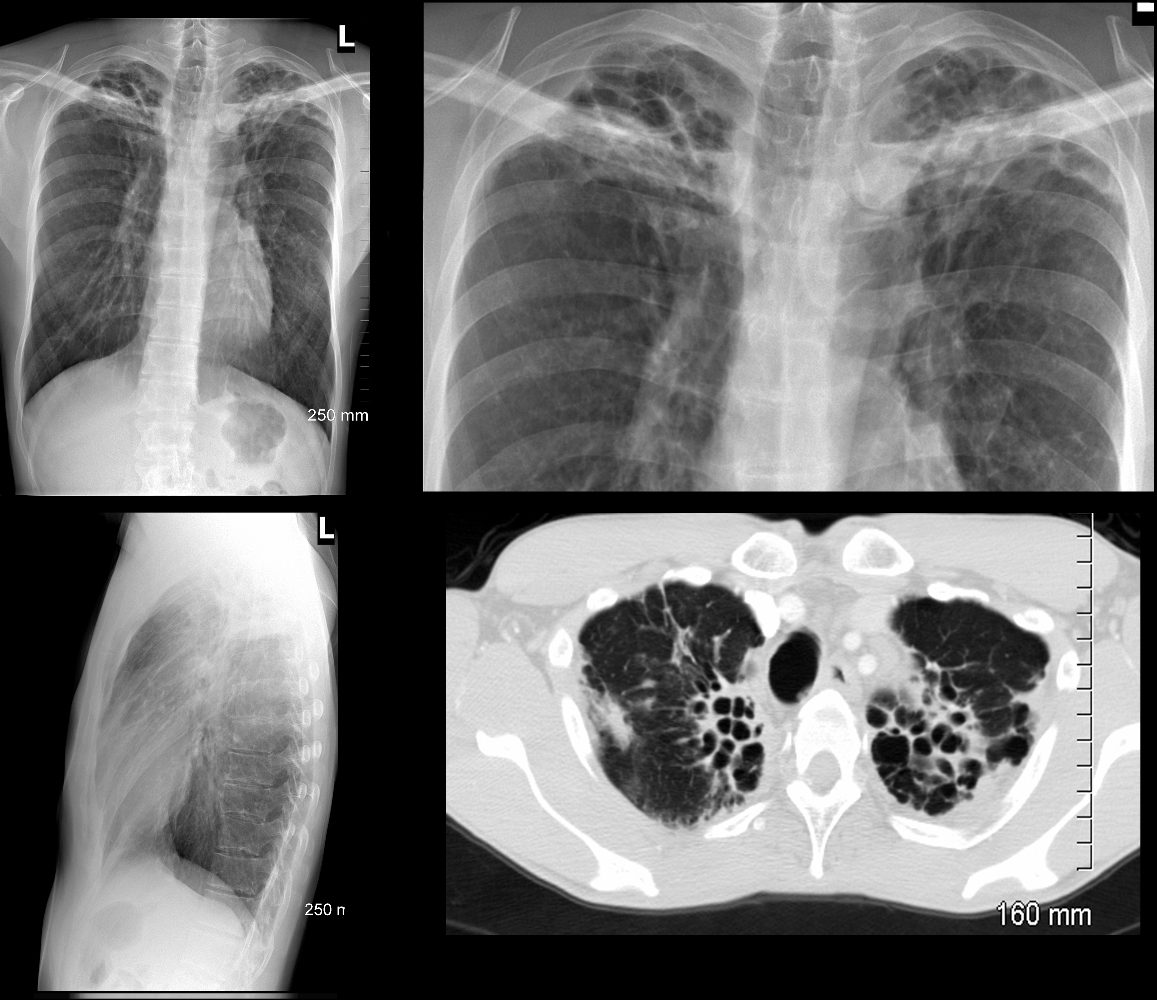
Progressive Bronchiectasis in the Apices

CXR and CT of a 53 year old male shows hyperinflation, bilateral hilar retraction and bronchiectasis in the apices bilaterally
Ashley Davidoff TheCommonvein.net
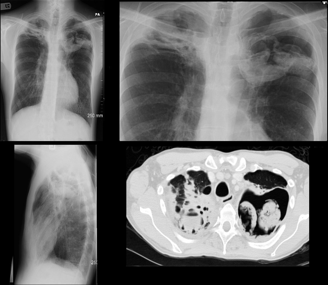
1Year Later Cavity with Aspergilloma in the left Apex and Consolidation and Abscess in the Right Apex

CXR and CT of a 54 year old male shows hyperinflation of the lungs, with large left apical cavity with aspergilloma and pneumonic consolidation and abscess formation in the apex of the right lung. These findings are consistent with chronic pulmonary aspergillosis
Ashley Davidoff TheCommonvein.net

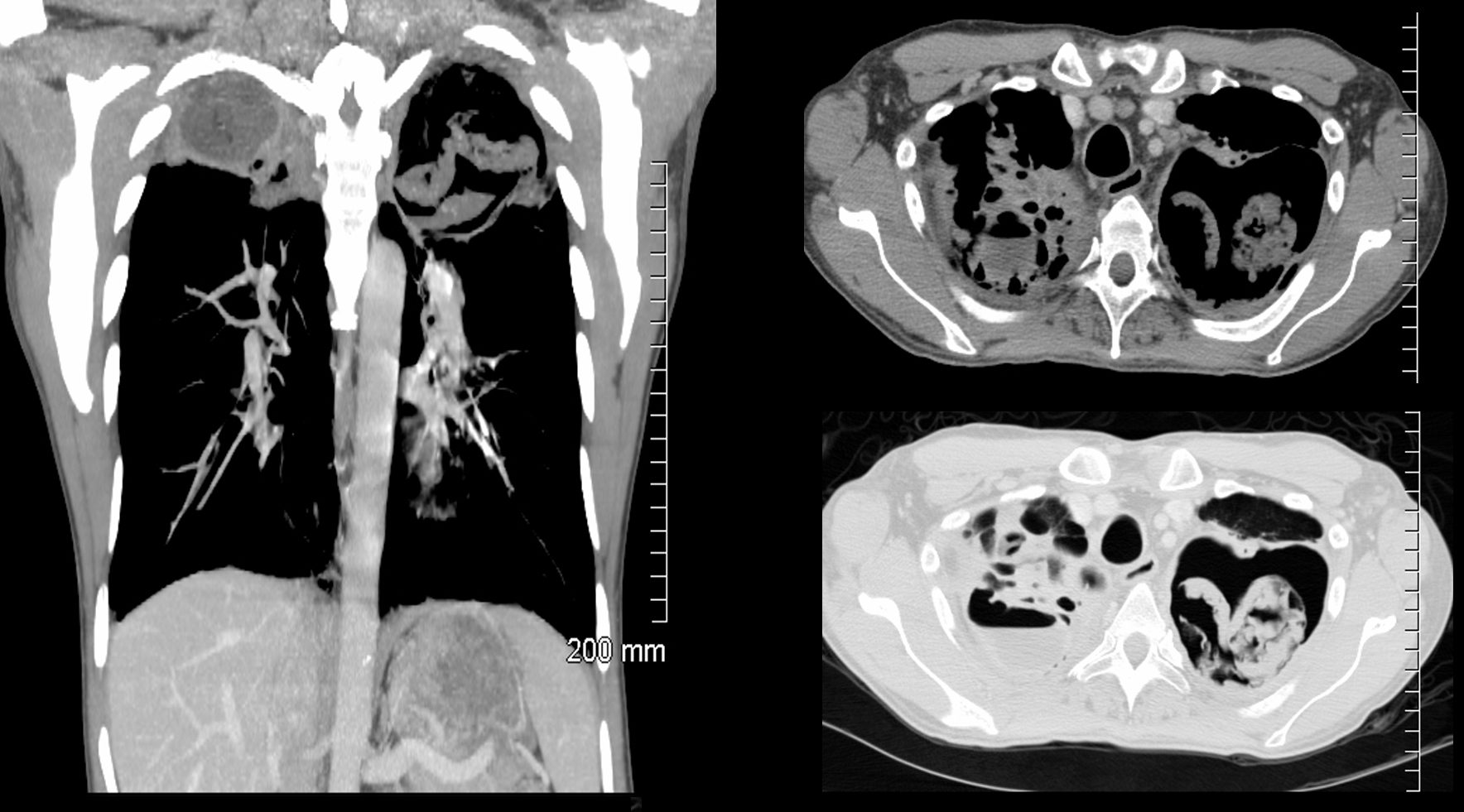
CT of a 54 year old male shows a large left apical cavity with aspergilloma. These findings are consistent with chronic pulmonary aspergillosis In the apex of the right lung, there is pneumonic consolidation and abscess formation . Note the air fluid level in the right apex on axial soft tissue and lung windows.
Ashley Davidoff TheCommonvein.net
Discussion
The patient presented 6 years prior and the CXR showed hyperinflation and findings consistent with latent TB characterized by hilar retraction and linear nodular changes in the apices. CT scan 4 years later showed progressive bronchiectasis and 1 year after that the left apex showed a large cavity suggesting secondary aspergillus infection with fungal ball . At the time serum and sputum tests suggested infection with aspergillus. The patient did have pulmonary symptoms with a wcc elevation likely from the lung abscess. It was felt the the aspergillus infection was likely of a chronic nature (Chronic Pulmonary Aspergillosis – CAP) The right apex showed consolidation and an air fluid indicating an abscess
