Pooja Sikka MD Ashley Davidoff MD
Problem
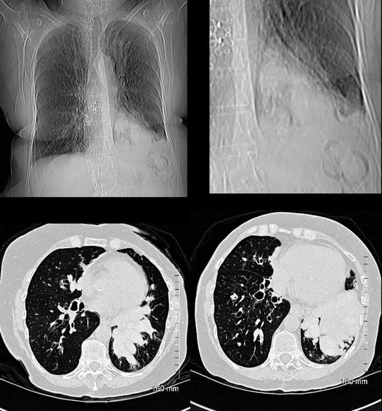
77 year old female with history of asthma, allergic bronchopulmonary aspergillosis (ABPA) and COPD
CT scout (a and magnified in b) shows lobular LLL infiltrate iof the lung Axial images show bilateral bronchiectatic airways . The LLL airways are more affected and are filled with mucus (finger in glove sign left lower image) becoming confluent and consolidative above the left hemidiaphragm right lower image).
Ashley Davidoff TheCommonVein.net
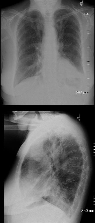
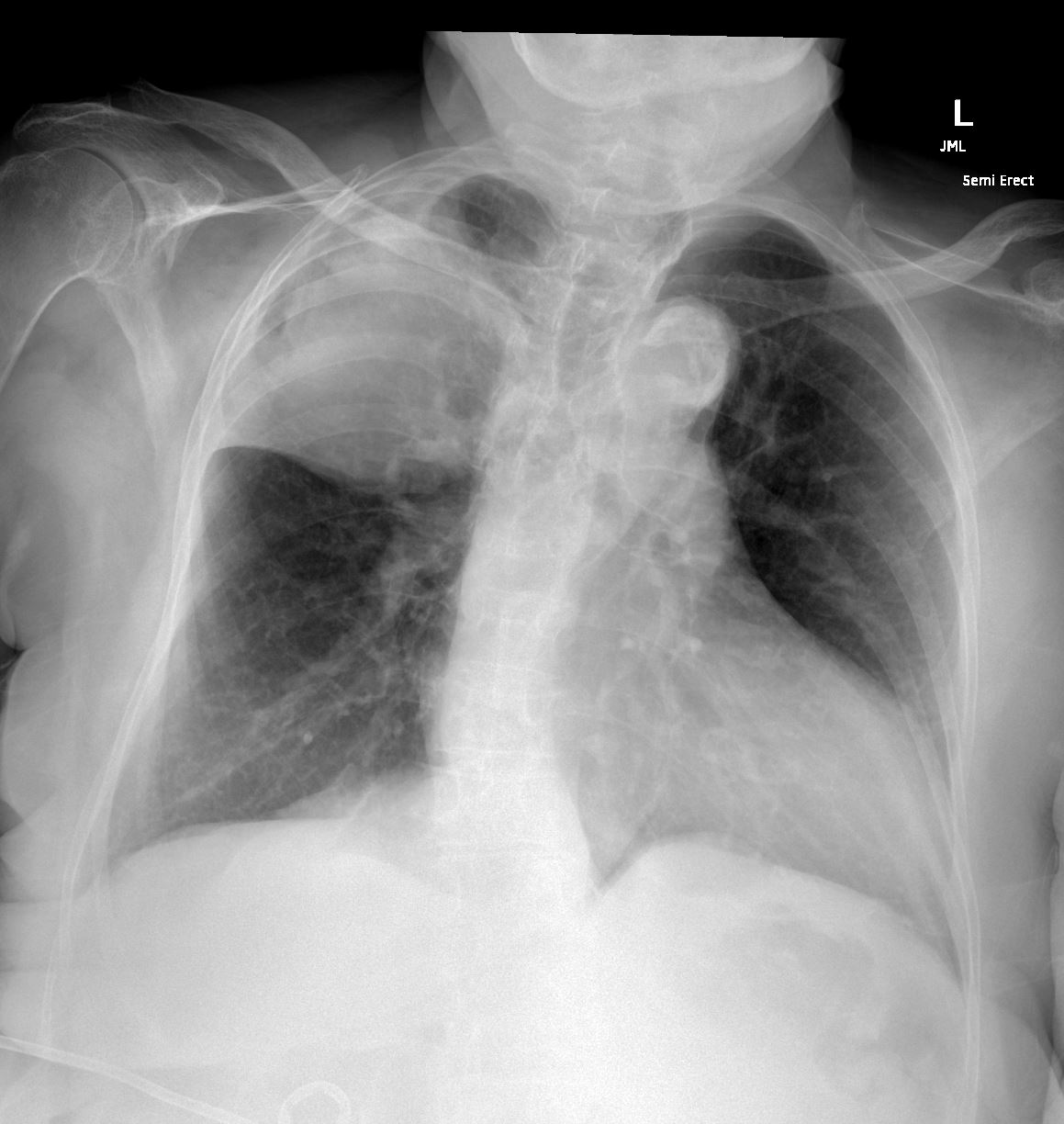
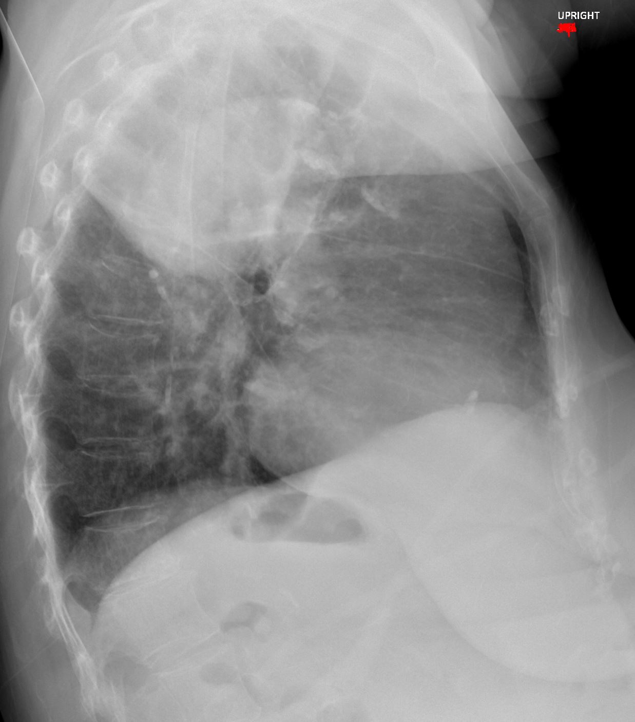
77 year old female with history of asthma, allergic bronchopulmonary aspergillosis (ABPA) and COPD
CXR shows hyperinflation, and consolidation in the left lower lobe silhouetting the left hemidiaphragm, with prominent bronchovascular bundles in the upper lung fields seen both on the PA and the lateral Diagnosis: Asthma Allergic Bronchopulmonary Aspergillosis (ABPA) COPD
Ashley Davidoff TheCommonVein.net
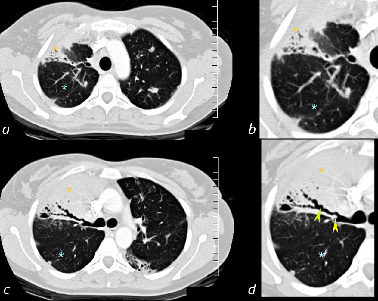
The axial images of the RUL are focused on the air filled varicoid bronchiectasis within the RUL atelectasis (lime green arrow in d) The shape of the posterior subsegmental airways has varicoid shape, and hence the terminology. The axial cuts on the left at 2 different levels, are magnified on the right images. The consolidation (yellow asterisk is noted in all 4 images – solid anteriorly, with the bronchiectatic posterior subsegmental airways posteriorly. There is a small focus of mucoid accumulation in the proximal segmental airway (yellow arrowhead d) The hyperinflated RLL is marked with a teal asterisk in all 4 images .
Ashley Davidoff MD TheCommonVein.net
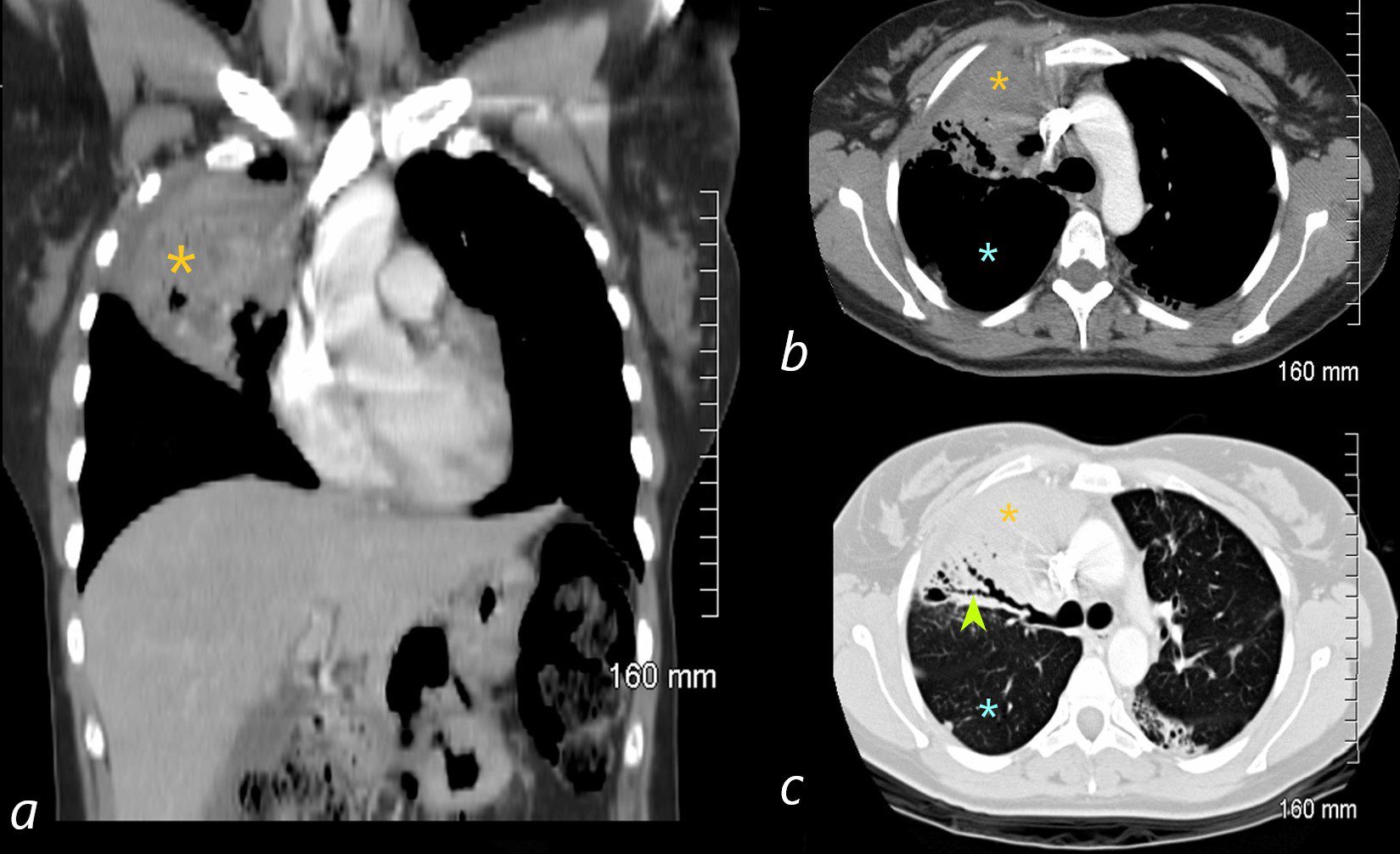
ABPA
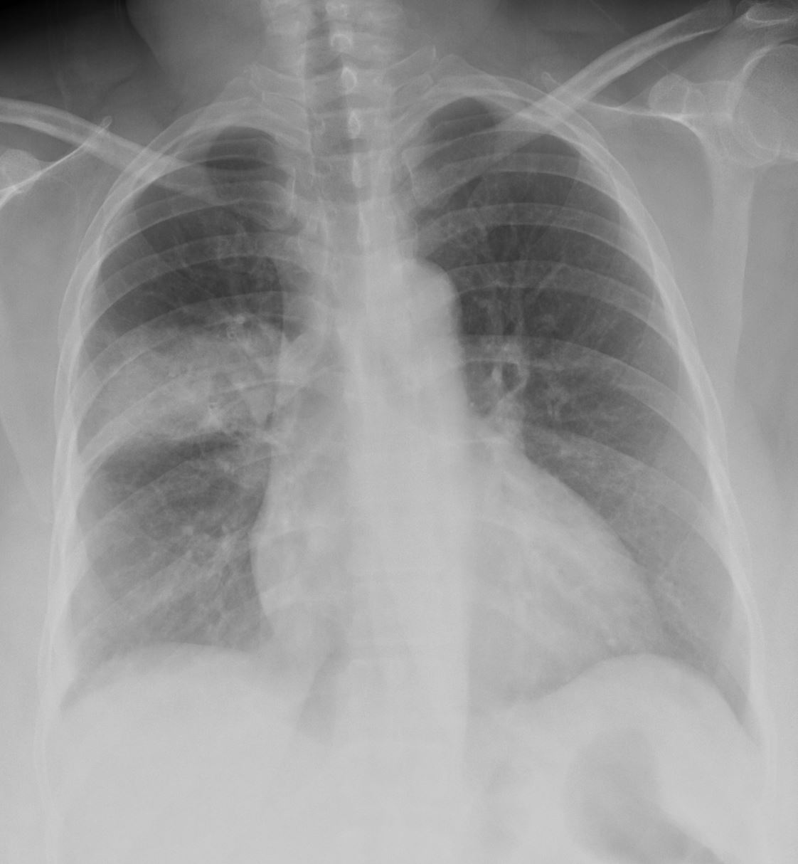
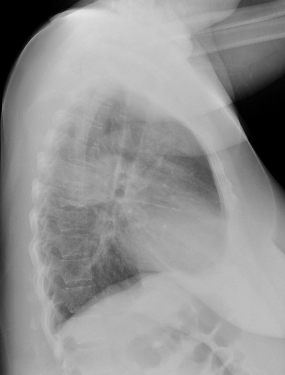
The coronal image is relatively anterior and hence presents as a dense consolidation of atelectasis (orange asterisk, a) In the axial images the hyperinflated RLL is seen posteriorly (teal asterisks in b and c) The region of varicose bronchiectasis is noted posteriorly (lime green arrow, c) When the net density of these 3 findings (consolidation, hyperinflated RLL and bronchiectasis in the LUL) are superimposed on CXR they present with a difficult interpretation since it is the overall net density that gets reflected. The CT scan helps us understand the findings
Ashley Davidoff MD TheCommonVein.net
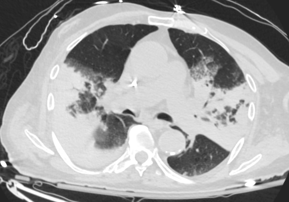
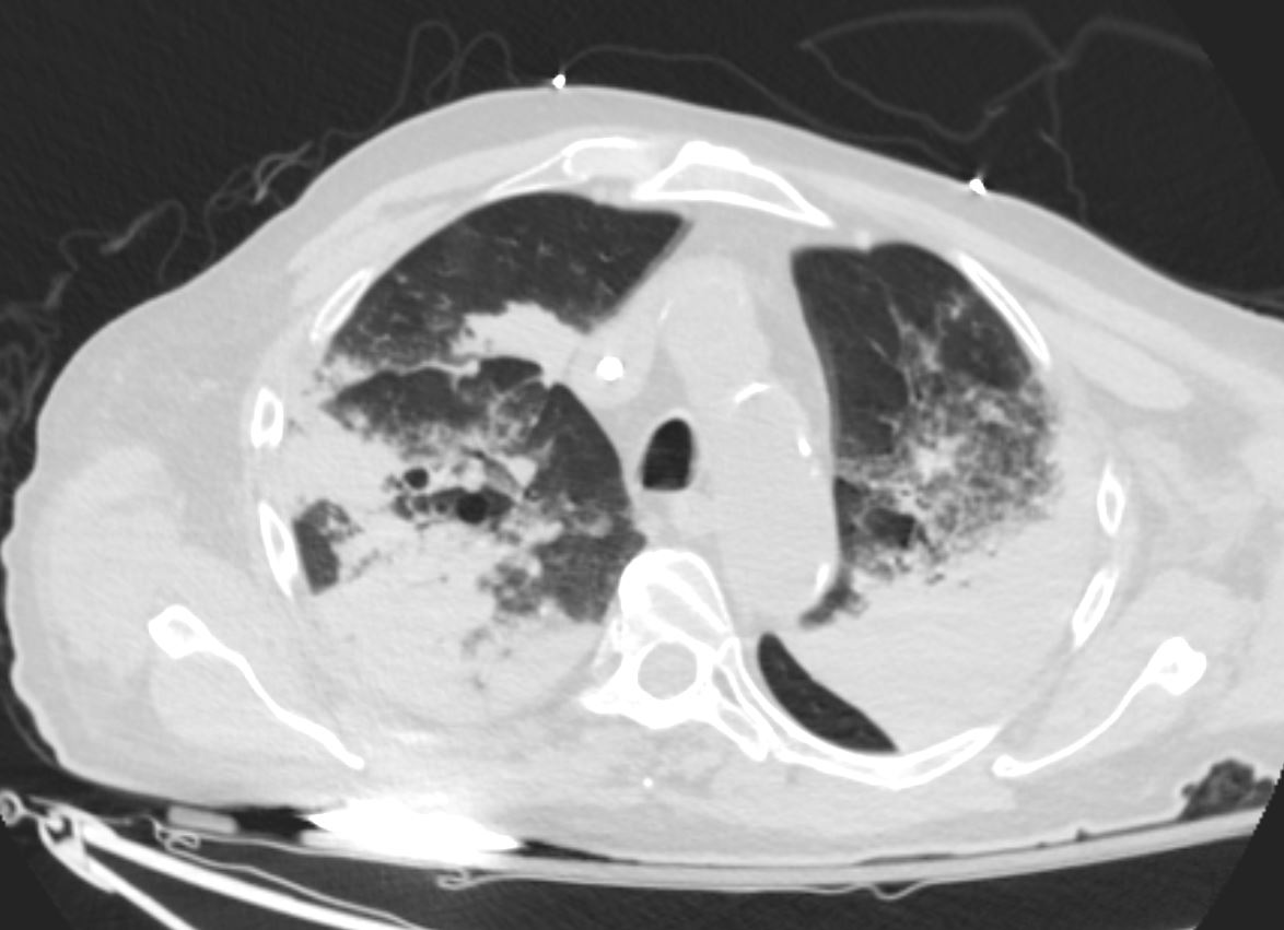
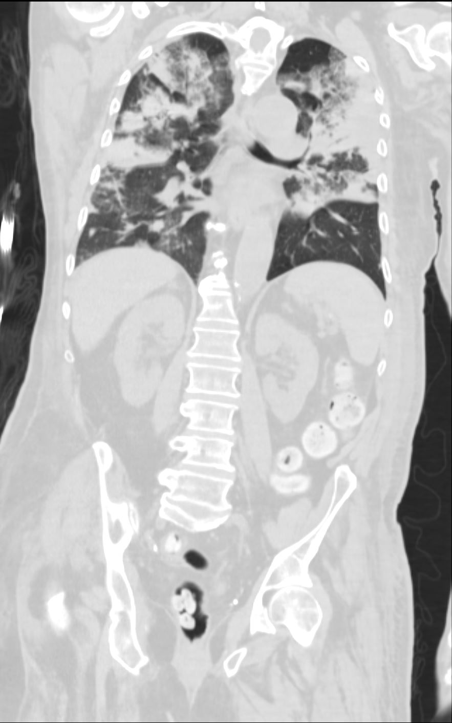
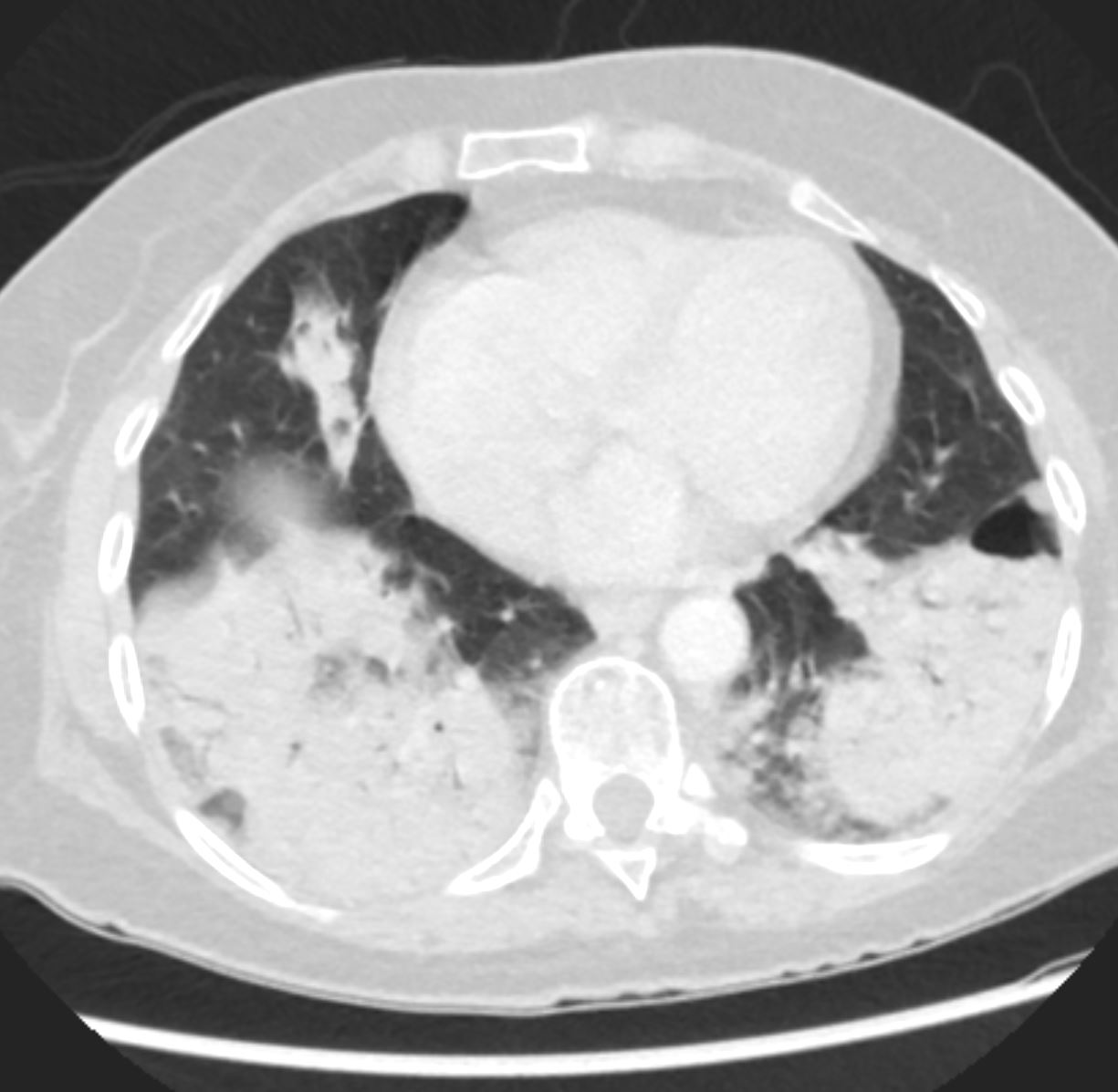
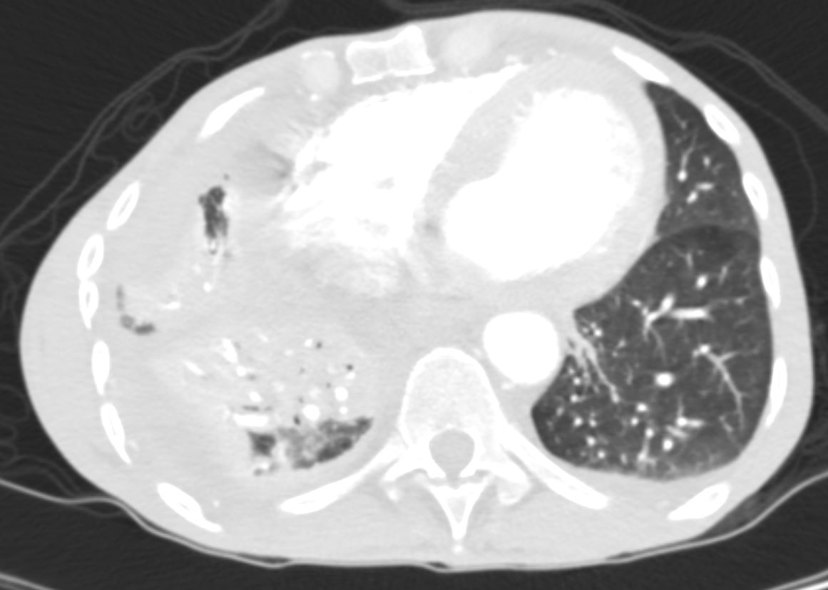
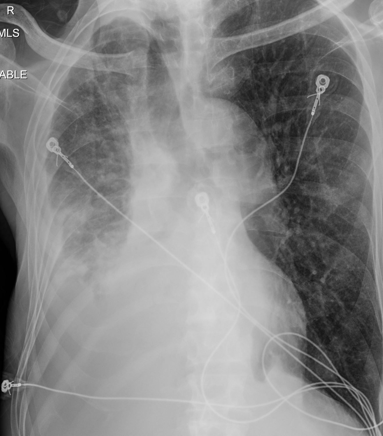
71 yo m w/ a hx of recently diagnosed ANCA-associated vasculitis (diagnosed by kidney biopsy), presents with acute respiratory failure. Bronchoscopic findings were consistent with diffuse alveolar hemorrhage associated with MSSA (Methicillin Sensitive Staph Aureus ) pneumonia/bacteremia. Note Consolidation surrounded by ground glass opacity, the latter likely reflecting a hemorrhagic component
CTscan shows multicentric consolidations likely a combination of alveolar hemorrhage and pneumonia
Ashley Davidoff TheCommonVein.net
Ashley Davidoff MD TheCommonVein.net
eosinophillic-pneumonia-003
Ashley Davidoff TheCommonVein.net Ashley Davidoff TheCommonVein.net RML RLL 002
Ashley Davidoff TheCommonVein.net Ashley Davidoff TheCommonVein.net RML RLL 001
Ashley Davidoff TheCommonVein.net RUL 47F 001
Ashley Davidoff TheCommonVein.net RUL 47F 002
Courtesy Ashley Davidoff TheCommonVein.net
Solution Next
Back to “Image First” Case List
