The frontal CXR shows subtle nodular changes in the right upper peripheral lung field (red circles) .
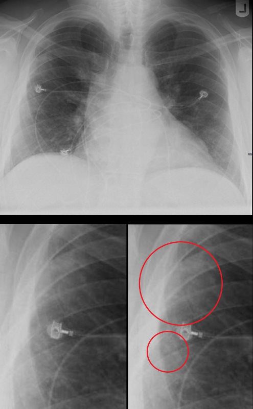
SARCOIDOSIS – CHARACTERISTIC NODULES
Ashley Davidoff MD
The CT examination scout film confirms 3 major regions of nodular change
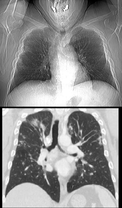
SARCOIDOSIS – CHARACTERISTIC NODULES
Ashley Davidoff MD
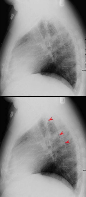
The lateral examination shows 3 regions of nodular changes (red arrowheads)
Ashley Davidoff MD
The Sagittal CT examination scout film confirms 3 major regions of nodular change in the posterior and superior segment of the RUL along the confluence of the right major and minor fissure and in the posterior segment of the left upper lobe peripherally.

Ashley Davidoff MD
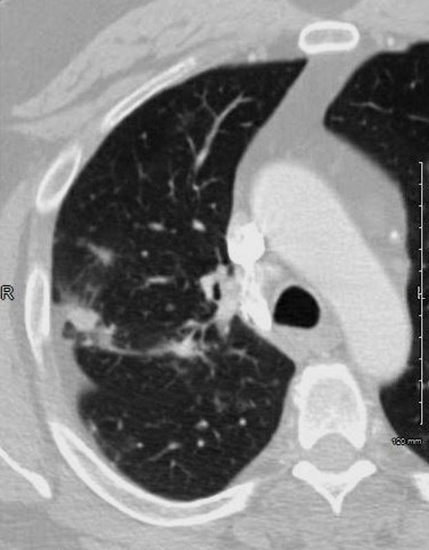
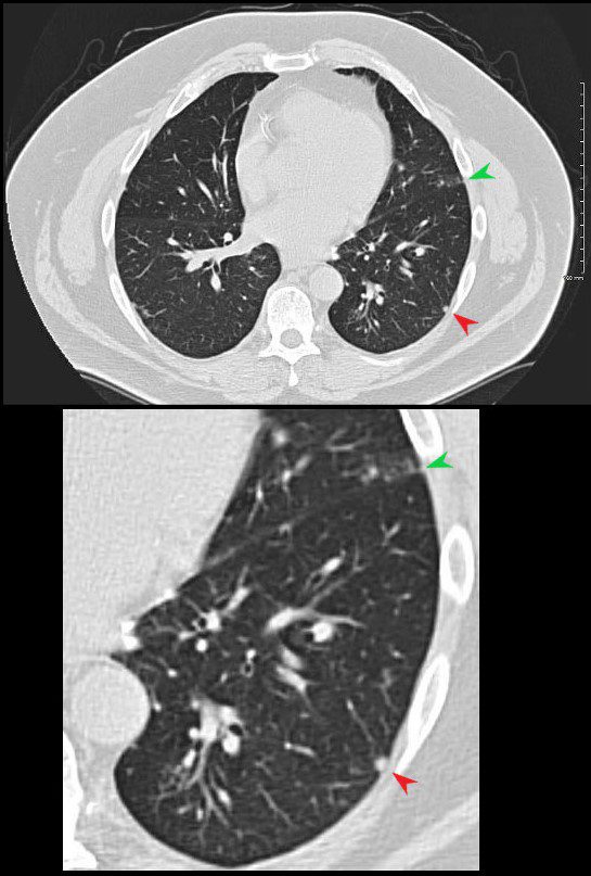
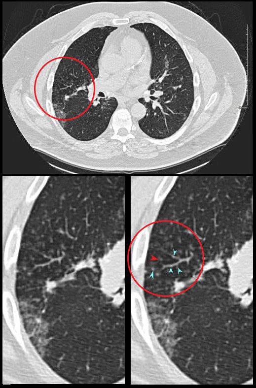
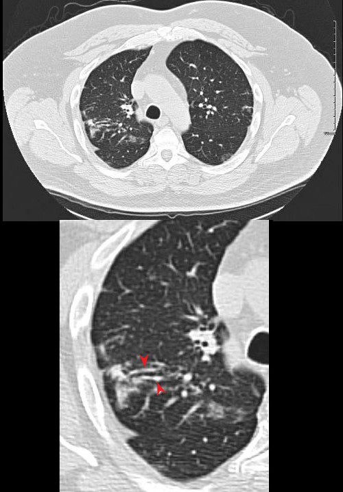
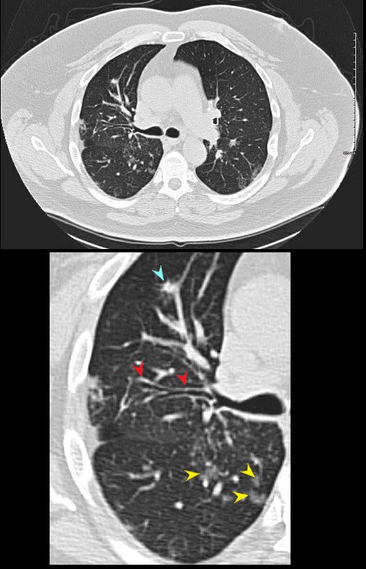
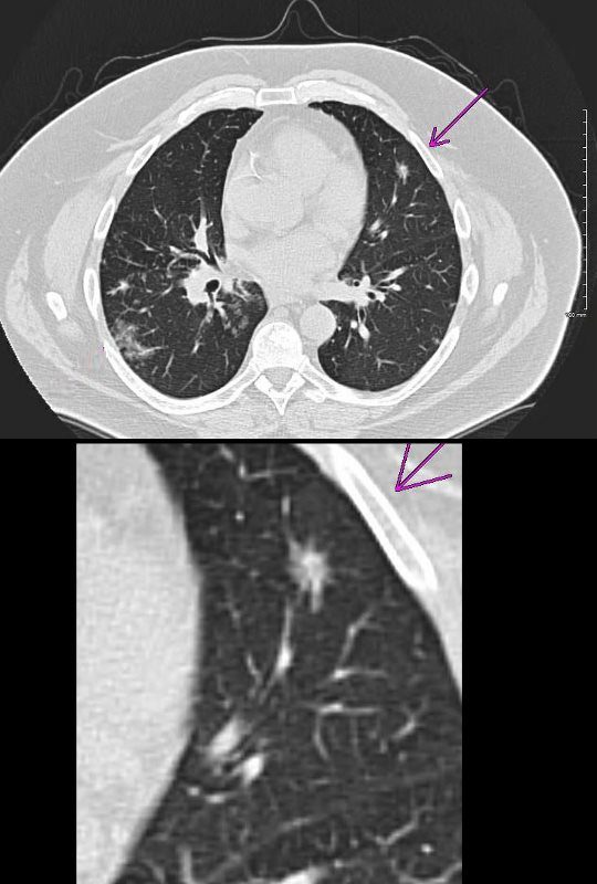
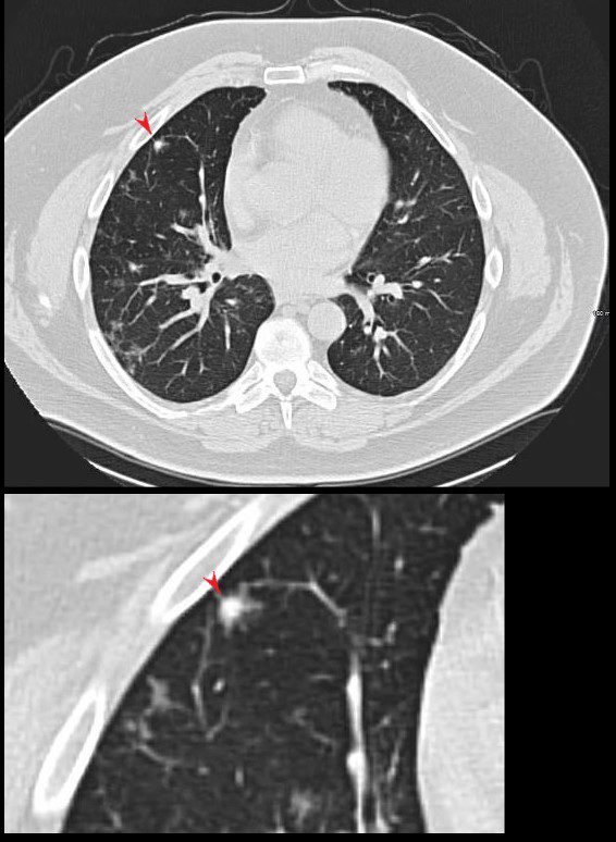
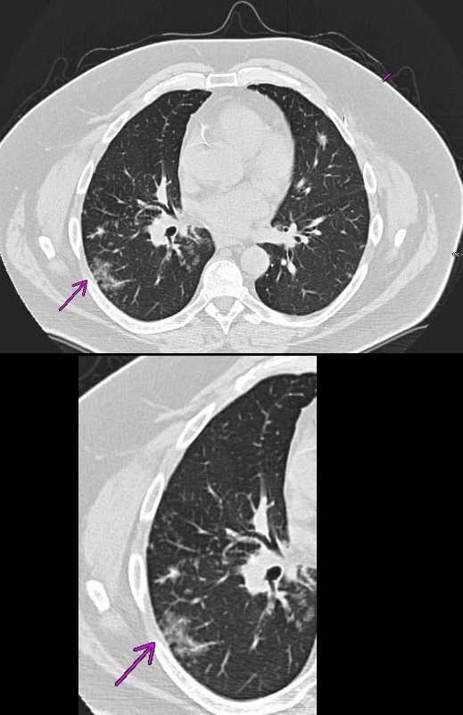
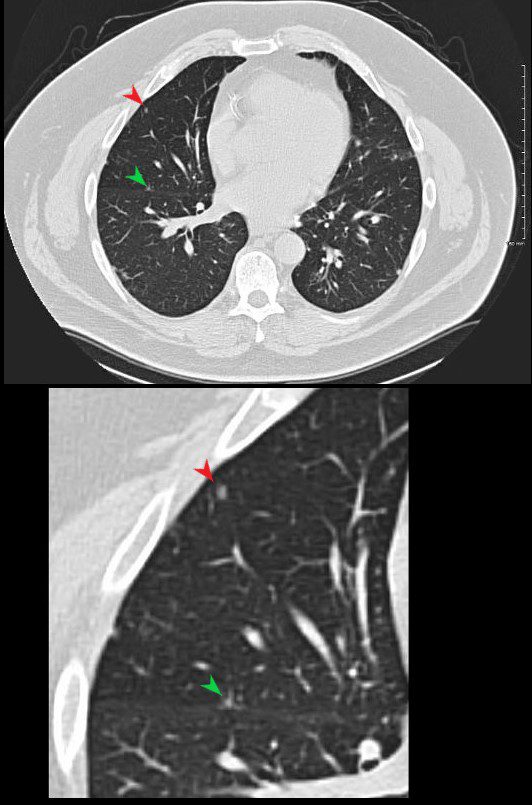
The axial images show a variety of characteristic changes including;
Ground glass opacity
Stellate or flame shaped nodules
Semi Solid nodules
Fissural based nodules
Subpleural nodules
Micronodules along the
lymphovascular and
bronchovascular bundles of the secondary lobule
Calcified nodule some of which are surrounded by soft tissue of the granuloma
Ashley Davidoff MD
Ashley Davidoff MD
Ashley Davidoff MD
Ashley Davidoff MD
Ashley Davidoff MD
Ashley Davidoff MD
Ashley Davidoff MD
Ashley Davidoff MD
Ashley Davidoff MD
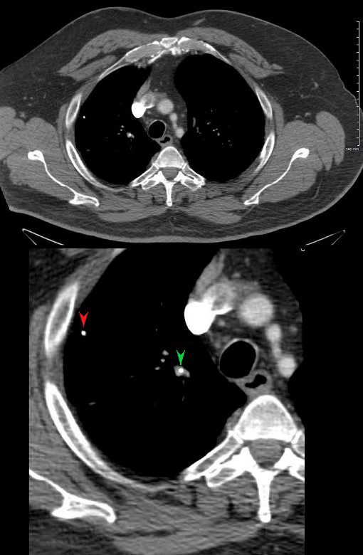
Ashley Davidoff MD
There are small calcified nodes in the mediastinum, but no significant pathologic adenopathy
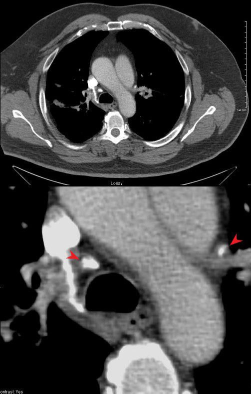
Ashley Davidoff MD
No obvious cardiac nor splenic involvement is noted
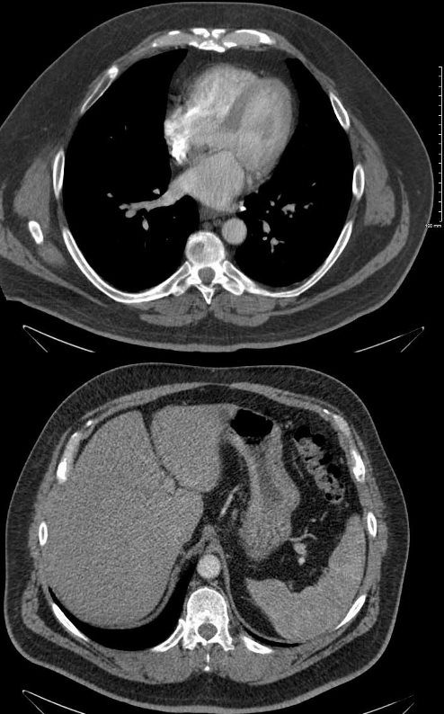
Ashley Davidoff MD
