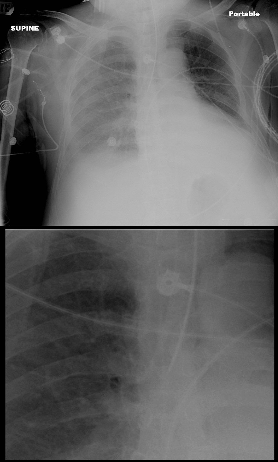
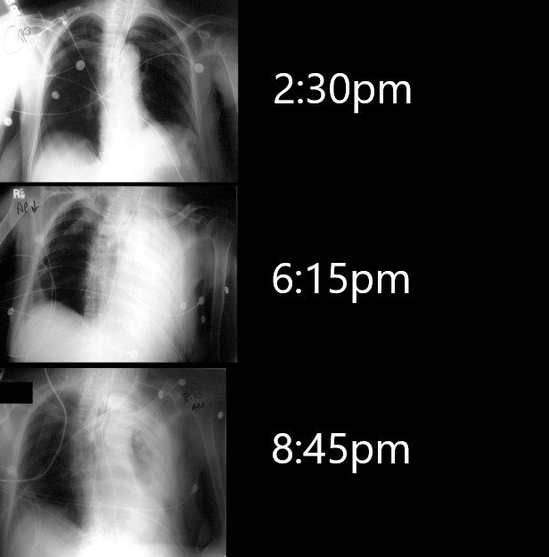
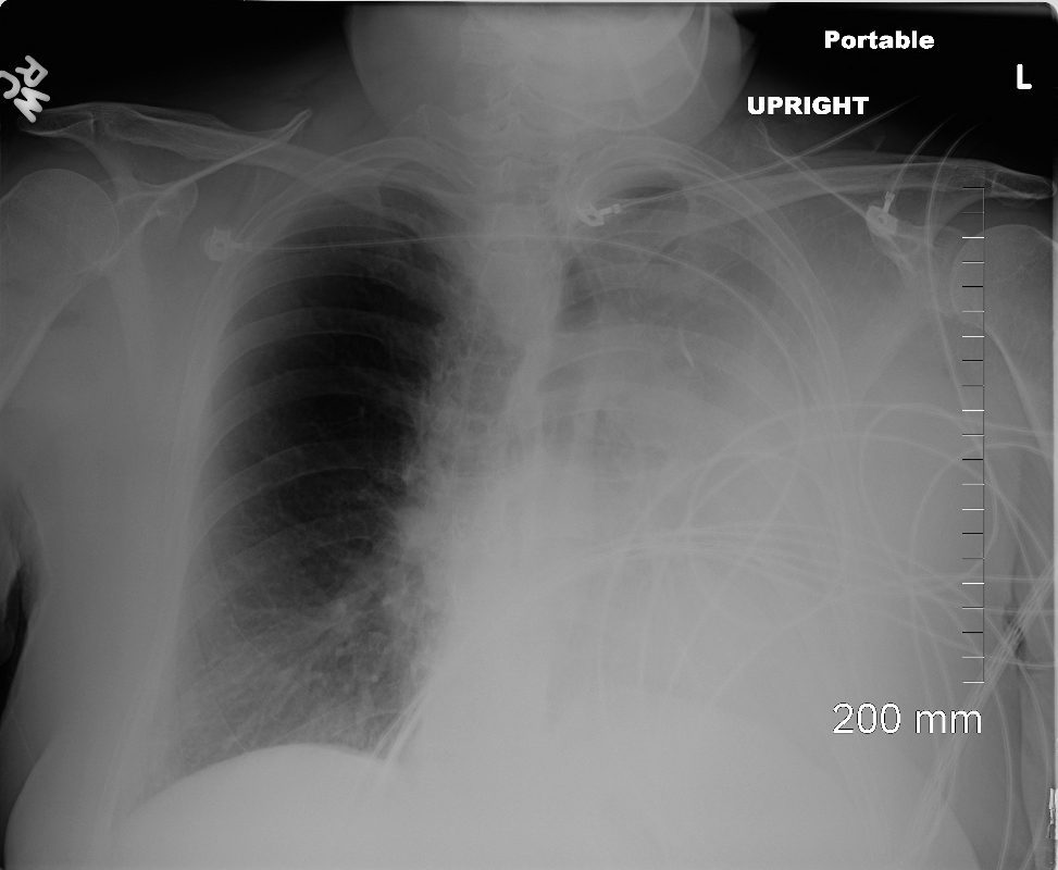
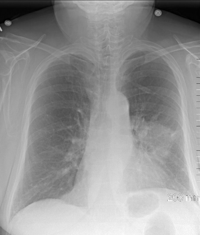
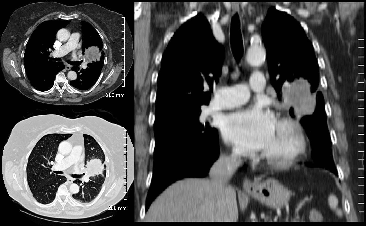
Faces of Atelectasis Time ETT in Right Main Stem Bronchus Post ETT in Right Main Stem BronchusPostObstructive Atelectasis of the left Lower LobeAshley Davidoff MD TheCommonVein.net42077 68 year old ICU patient has an Xray at 2:30pm to check tube position . 4 hours later he presents with a acute respirtatory difficulty and the re is a white out of the left lung (middle image). After suction, a repeat CXR approximately 2 hours later shows improving aeration. The rapid response to suction usually infers obstruction from a mucus plug or inhalation of a foreign body with removal . Ashley Davidoff MD TheCommonVein.net 70231cL White Out After bronchoscopy for Central Cancer 72 year old female with left upper lobe mass s/p bronchoscopy complicated by lung collapse characterised by consolidation, elevated left hemidiaphragm and left ward shift of the cardio- mediastinal shadowAshley Davidoff MD TheCommonVein.net 1 Day After Bronchoscopy Re- Aeration 72 year old female with left upper lobe mass 1 day after bronchoscopy which was complicated by lung collapse (atelectasis). The CXR has returned to baseline with left hilar massAshley Davidoff MD TheCommonVein.net Atelectasis Following Bronchoscopy72 year old female presents with a cough. CT shows a suspicious 3.2cms central, spiculated left upper lobe mass.Ashley Davidoff MD TheCommonVein.net Links and References TCV Introduction to Atelectasis Faces of Atelectasis (Diseases) Size Shape Position Character Time Types Compressive Atelectasis Post Obstructive Atelectasis Rounded Atelectasis Cicatrization Atelectasis Adhesive Atelectasis Atelectasis Cases