CXR Pectus Excavatum
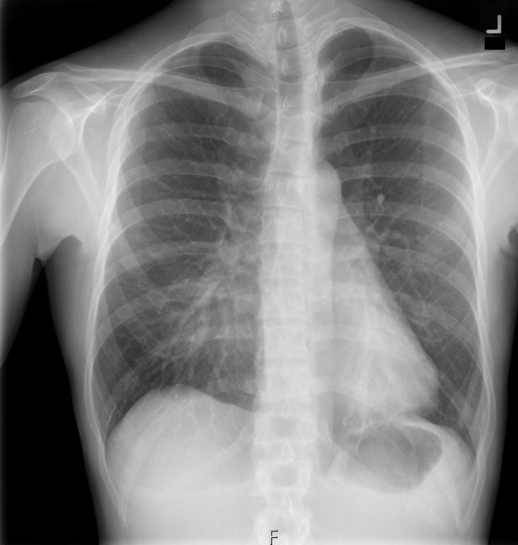
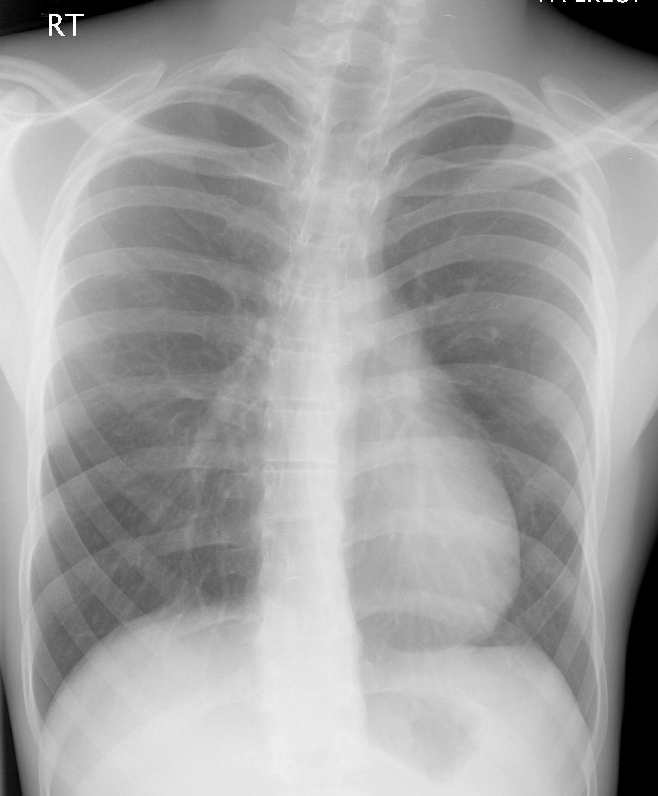
33-year-old female presents with a cough. Chest X-ray in the frontal view shows a region of increased density in the medial right lower lung field. The cardio mediastinal shadow is shifted to the left. These findings are consistent with pectus excavatum.
Courtesy Ashley Davidoff MD TheCommonVein.net 136533a
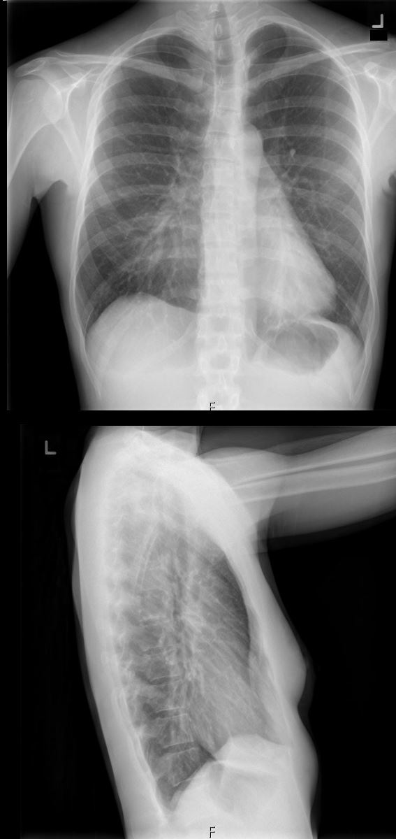
33-year-old female presents with a cough. Chest X-ray in the frontal view shows a region of increased density in the medial right lower lung field. The cardio mediastinal shadow is shifted to the left. On the lateral view a moderate sized pectus excavatum causes a decrease in the A_P diameter of the chest, compresses the lung accounting for the increased density and causes the cardio-mediastinal shadow to shift leftward.
Courtesy Ashley Davidoff MD TheCommonVein.net 136533b
68-year-old female presents with a severe pectus excavatum.
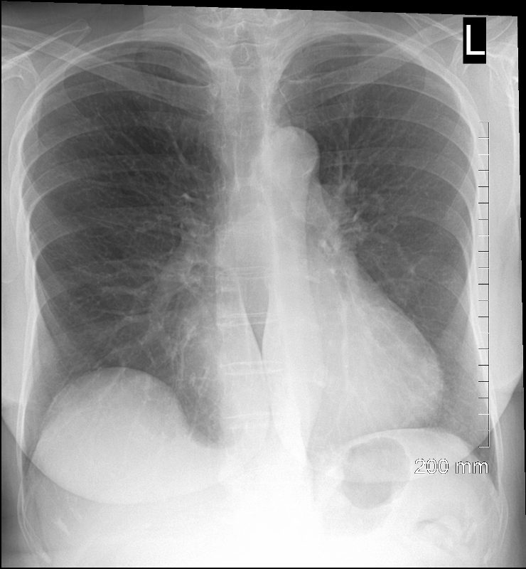
68-year-old female presents with a severe pectus excavatum. CXR in the frontal view shows horizontal orientation of the ribs and distortion of the right heart border
Ashley Davidoff MD TheCommonVein.net 270Lu 121391
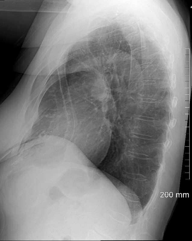
68-year-old female presents with a severe pectus excavatum. CXR in the lateral view shows significant depression of the sternum
Ashley Davidoff MD TheCommonVein.net 270Lu 121392
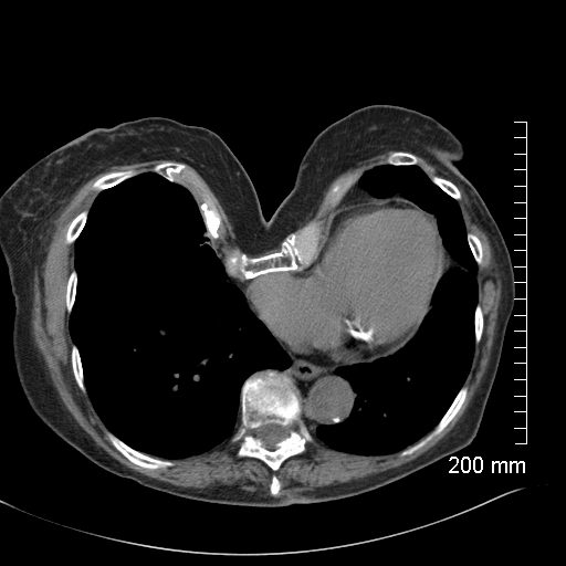
68-year-old female presents with severe pectus excavatum. CT in the axial plain shows significant depression of the sternum, and dextrocardia
Ashley Davidoff MD TheCommonVein.net 270Lu 121393
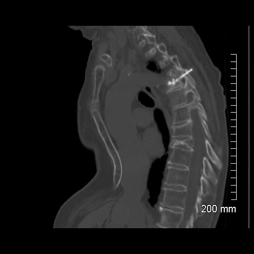
68-year-old female presents with severe pectus excavatum. CT in the sagittal plain shows significant depression of the lower sternum, compression fracture of a midthoracic vertebra and surgical hardware in a proximal vertebra
Ashley Davidoff MD TheCommonVein.net 270Lu 121396
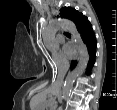
68-year-old female presents with severe pectus excavatum. CT in the sagittal plain shows significant depression of the lower sternum,
Ashley Davidoff MD TheCommonVein.net 270Lu 121399
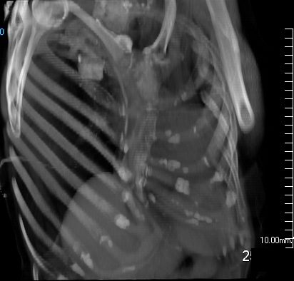
68-year-old female presents with severe pectus excavatum. 3D CT in the RAO projection shows significant depression of the sternum
Ashley Davidoff MD TheCommonVein.net 270Lu 121397
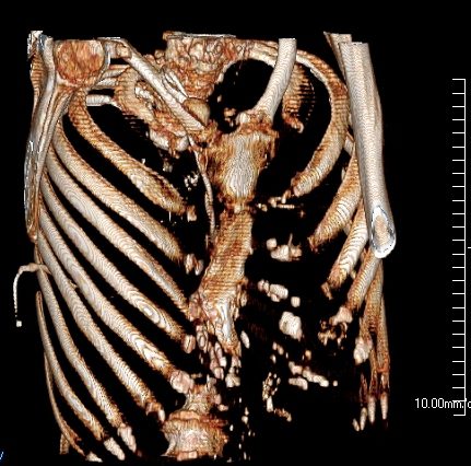
68-year-old female presents with severe pectus excavatum. 3D CT in the RAO projection shows significant depression of the sternum
Ashley Davidoff MD TheCommonVein.net 270Lu 121398
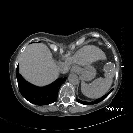
68-year-old female presents with severe pectus excavatum. CT in the axial plain shows significant depression of the region of the xiphi-sternum. Noted calcified cystic lesion of the spleen
Ashley Davidoff MD TheCommonVein.net 260Lu 21395
Pectus Excavatum
CXR
Ashley Davidoff MD
thecommonvein.net
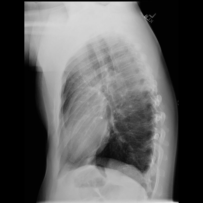
This a normal lateral chest X ray of a 40 year old male with mild pectus excavatum
Ashley Davidoff MD
