A bleb in radiology refers to a small, thin-walled air-filled space located within or just under the visceral pleura of the lung. Blebs are typically less than 1-2 cm in diameter and are formed due to the rupture or dissection of alveoli into the subpleural space. They are benign but can predispose to complications like spontaneous pneumothorax if they rupture.
Radiological Features
- Chest X-Ray (CXR):
- Often not visible due to their small size and thin walls.
- Indirect signs, such as a pneumothorax, may suggest the presence of a ruptured bleb.
- Occasionally seen as a small, peripheral air-filled lucency near the lung apex.
- Computed Tomography (CT):
- Appearance:
- Thin-walled, round, or irregular air-filled space located just beneath the visceral pleura.
- Typically subpleural and most commonly found in the apices of the upper lobes.
- Size:
- Generally less than 2 cm in diameter.
- Associated Findings:
- Adjacent emphysematous changes (e.g., bullae, paraseptal emphysema).
- Pneumothorax if the bleb has ruptured.
- Appearance:
- Ultrasound:
- Rarely used for bleb detection but may help identify pneumothorax caused by a ruptured bleb through the absence of lung sliding or presence of a “lung point.”
Common Causes and Associations
Blebs can occur idiopathically or secondary to underlying lung conditions:
- Idiopathic (Primary):
- Often seen in young, tall, thin males with no underlying lung disease.
- Common cause of primary spontaneous pneumothorax.
- Secondary to Lung Pathology:
- Paraseptal Emphysema:
- Destruction of alveolar walls near the pleura predisposes to bleb formation.
- Chronic Obstructive Pulmonary Disease (COPD):
- Increased intrapulmonary pressures can lead to bleb formation.
- Cystic Lung Diseases:
- Rare conditions like lymphangioleiomyomatosis (LAM) or Birt-Hogg-Dubé syndrome may present with blebs.
- Paraseptal Emphysema:
- Trauma or Mechanical Ventilation:
- Barotrauma can lead to the formation or rupture of blebs.
Differential Diagnosis
- Bullae:
- Larger than blebs (>2 cm), with thicker walls, and located deeper within the lung parenchyma.
- Cysts:
- Well-defined, air-filled spaces with thicker walls and typically associated with specific lung diseases (e.g., LAM, histiocytosis X).
- Cavities:
- Air-filled spaces with thick, irregular walls due to infection, malignancy, or necrosis.
- Pneumatocele:
- Air-filled spaces with thin walls caused by acute lung injury or infection.
Complications
- Spontaneous Pneumothorax:
- A ruptured bleb allows air to escape into the pleural space, leading to lung collapse.
- Recurrent Pneumothorax:
- Patients with multiple or persistent blebs are at risk of recurrence.
- Tension Pneumothorax:
- Rare but life-threatening if rupture results in air trapping under pressure.
Management
- Observation:
- Small, asymptomatic blebs often require no intervention.
- Pneumothorax Treatment:
- Chest tube placement or needle decompression for large or tension pneumothorax.
- Surgical Options:
- Video-Assisted Thoracoscopic Surgery (VATS):
- Used to remove blebs and perform pleurodesis in recurrent cases.
- Pleurodesis (chemical or mechanical) to prevent recurrence.
- Video-Assisted Thoracoscopic Surgery (VATS):
Clinical Relevance
Identifying blebs on imaging is critical for managing conditions like spontaneous pneumothorax and differentiating blebs from other cystic or air-filled lesions of the lung. CT is the gold standard for detecting and characterizing blebs due to its superior resolution.

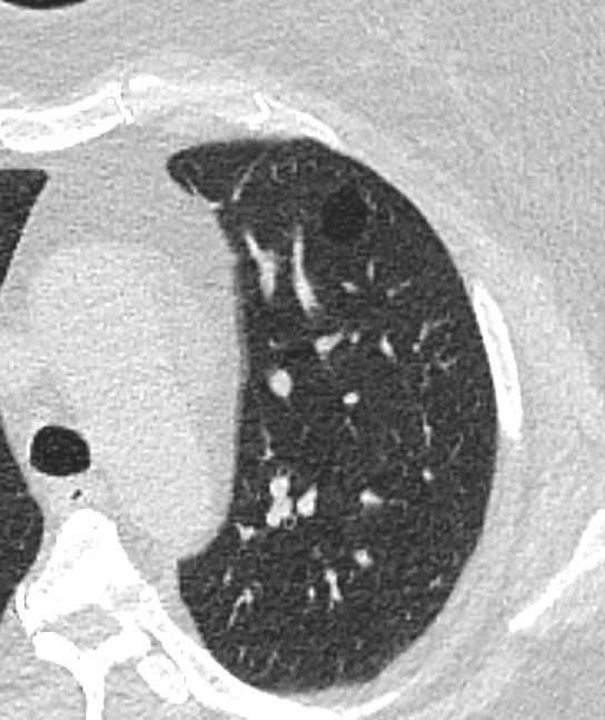
CT scan in the axial plain in a 59 Year old female with emphysema, shows a 1.6cms bleb in the anterior segment of the left upper lobe
Ashley Davidoff MD TheCommonVein.net 136619cL

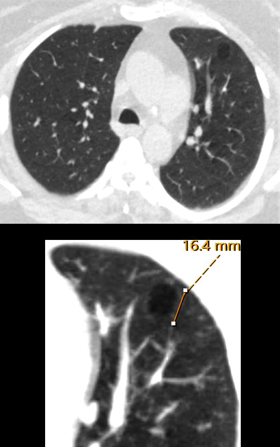
CT scan in the axial and coronal plains in a 50 year old female shows a 1.cms bleb in the apex of the left upper lobe
Ashley Davidoff MD TheCommonVein.net 137860

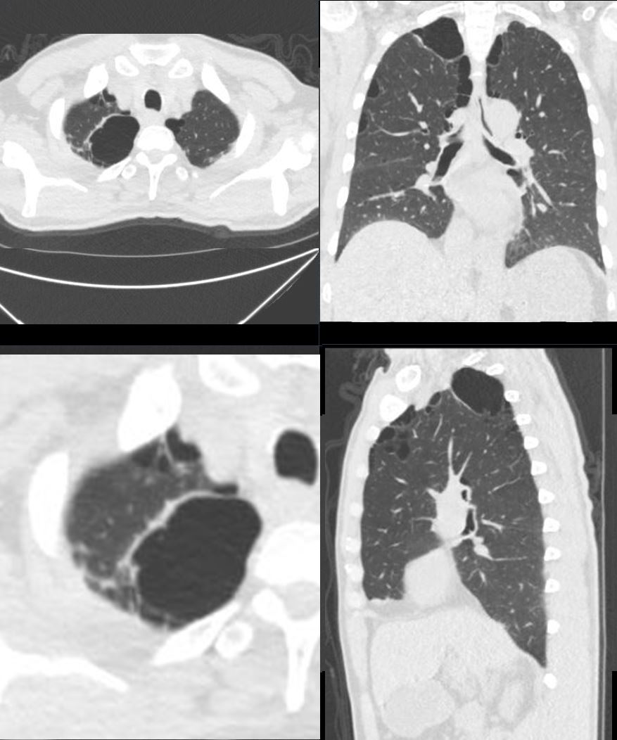
CT scan in the axial and coronal plains in a 55 year old male shows a combination of blebs bulla and paraseptal emphysema most prominent in the apex of the right upper lobe
Ashley Davidoff MD TheCommonVein.net 136929
Links and References
Fleischner Society
Anatomy.—A bleb is a small gas-containing space within the visceral pleura or in the subpleural lung, not larger than 1 cm in diameter (,25).
CT scans.—A bleb appears as a thin-walled cystic air space contiguous with the pleura. Because the arbitrary (size) distinction between a bleb and bulla is of little clinical importance, the use of this term by radiologists is discouraged.

