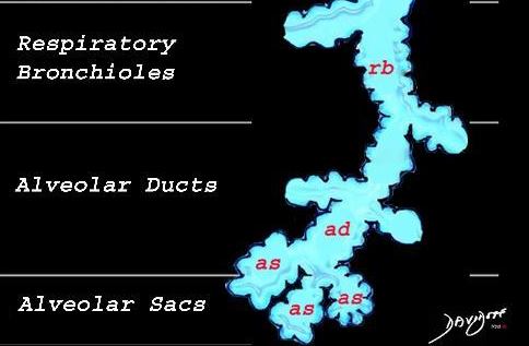
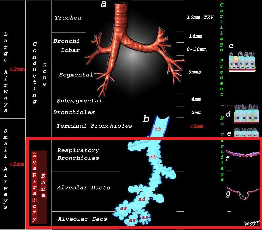
Airspace in the lungs or respiratory zone, refers to the alveolar region where gas exchange occurs, including the alveoli, alveolar ducts, and respiratory bronchioles. Pathologically, the term “airspace disease” is used to describe conditions where the airspaces become filled with substances such as fluid, blood, pus, or cells, as seen in pneumonia, pulmonary edema, or hemorrhage, which impairs gas exchange. As a result, lung function is compromised, leading to symptoms like cough, dyspnea, and hypoxemia. Diagnosis of airspace disease involves clinical evaluation, imaging studies such as chest X-rays or CT scans that show opacification (e.g., consolidation or ground-glass opacity), and laboratory tests depending on the underlying cause, such as sputum cultures or blood tests for infection or inflammation.

The respiratory zone or airspace refers to the alveolar region where gas exchange occurs, including the respiratory bronchioles, alveolar ducts, alveolar sacs, and alveoli.
Courtesy Ashley Davidoff MD TheCommonVein.net

The respiratory zone or airspace outlined in red refers to the alveolar region where gas exchange occurs, including the respiratory bronchioles, alveolar ducts, alveolar sacs, and alveoli.
Courtesy Ashley Davidoff MD TheCommonVein.net
Fleischner Society
airspace
Anatomy.—An airspace is the gas-containing part of the lung, including the respiratory bronchioles but excluding purely conducting airways, such as terminal bronchioles.
Radiographs and CT scans.—This term is used in conjunction with consolidation, opacity, and nodules to designate the filling of airspaces with the products of disease (,14).
