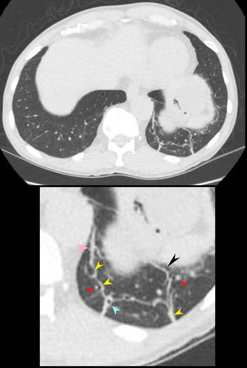
Architectural distortion refers to an abnormality in the structure or organization of lung tissue. This appearance typically involves disruption of the normal arrangement of pulmonary vessels, bronchi, and surrounding structures, giving a “pulled” or “warped” look to the lung parenchyma. Architectural distortion is a concerning finding, as it suggests that there is scarring, fibrosis, or a mass—that is disrupting the normal lung architecture.

CT scan through the lower lung field reveal findings consistent with architectural distortion. The normal arrangement of pulmonary vessels, bronchi, and surrounding structures in the left lower lobe, have a “pulled” or “warped” appearance to the lung parenchyma. In this case scarring associated with bronchial disease with thickening of the bronchial wall, thickening of the interlobular septa, and the presence of and centrilobular nodules, results in linear atelectasis and linear subpleural bands with distortion of the architecture
Ashley Davidoff MD TheCommonVein.net 136787-01

CT scan through the lower lung field reveal findings consistent with architectural distortion. The normal arrangement of pulmonary vessels, bronchi, and surrounding structures in the left lower lobe, have a “pulled” or “warped” appearance to the lung parenchyma (black arrowhead). In this case scarring associated with bronchial disease with thickening of the bronchial wall (teal arrowhead), thickening of the interlobular septa, (yellow arrowheads), and the presence of centrilobular nodules (red arrowheads) results in linear atelectasis and linear subpleural bands (pink arrowhead) with distortion of the architecture
Ashley Davidoff MD TheCommonVein.net 136787-01 136787-01L

CT scan through the lower lung field reveal findings consistent with architectural distortion. The normal arrangement of pulmonary vessels, bronchi, and surrounding structures in the left lower lobe, have a “pulled” or “warped” appearance to the lung parenchyma. In this case scarring associated with bronchial disease with thickening of the bronchial wall, thickening of the interlobular septa, and the presence of and centrilobular nodules, results in linear atelectasis and linear subpleural bands with distortion of the architecture
Ashley Davidoff MD TheCommonVein.net 136787-02
Causes of Architectural Distortion
Architectural distortion can be caused by various conditions, including:
- Pulmonary fibrosis or interstitial lung disease, where chronic inflammation or scarring alters lung structure.
- Post-infectious scarring, such as after tuberculosis or bacterial pneumonia.
- Tumors (primary or metastatic), which can “pull” surrounding structures and alter the lung architecture.
- Prior lung surgery or trauma, which can lead to scarring and distortion in the affected areas.
- Granulomatous diseases like sarcoidosis, which can produce fibrotic changes.
Radiologic Features
On imaging (X-ray or CT scan), architectural distortion may present with:
- Displacement or distortion of bronchi and pulmonary vessels.
- Retraction of pleura or fissures towards an area of fibrosis or scarring.
- Areas of dense, irregular tissue that lack the typical open and airy appearance of normal lung tissue.
Clinical Significance
Architectural distortion is a non-specific finding but often indicates underlying chronic disease, fibrosis, or malignancy. In cases where architectural distortion is present, additional imaging (e.g., high-resolution CT) or even biopsy may be required to identify the underlying cause, especially if there is no prior history of infection, surgery, or known lung disease.
Architectural distortion is thus an important radiologic sign, prompting further evaluation to rule out serious conditions, including malignancy or advanced interstitial lung disease.
Fleischner Society
Pathology.—Architectural distortion is characterized by abnormal displacement of bronchi, vessels, fissures, or septa caused by diffuse or localized lung disease, particularly interstitial fibrosis.
CT scans.—Lung anatomy has a distorted appearance and is usually associated with pulmonary fibrosis (,Fig 7) and accompanied by volume loss.
