Parts
Size
Shape
Position
Character
Time Associated Findings
Infection
Inflammation
Malignancy
Malignant Pericarditis

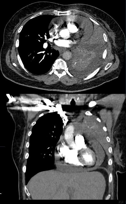
62-year-old female presents with acute dyspnea and chest pain
CT in the axial and coronal plane shows a small to moderate pericardial effusion, left lung collapse and small pleural effusion.
Echocardiogram revealed increased right sided pressures but no “frank” tamponade, and was subsequently drained
Ashley Davidoff MD TheCommonVein.net 298Lu 136725

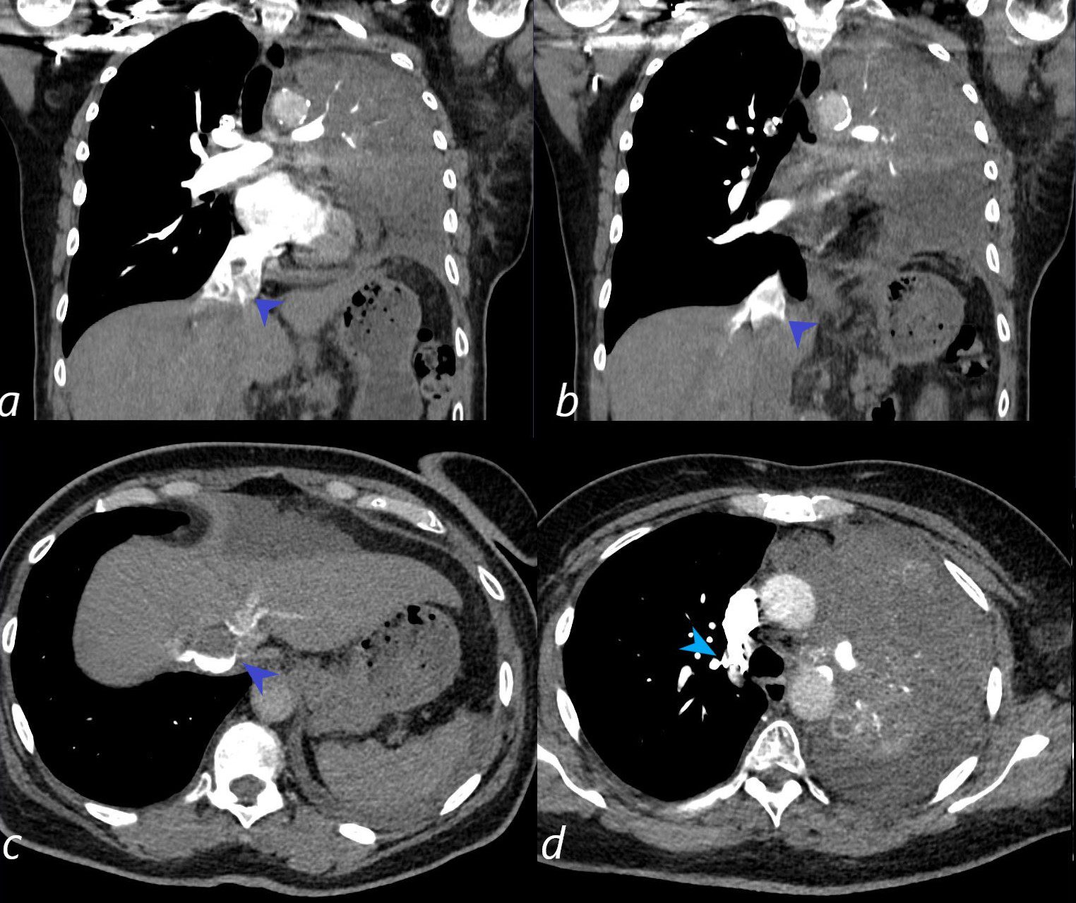
62-year-old female presents with acute dyspnea and chest pain
CT in the coronal and axial planes shows a small to moderate pericardial effusion, left lung collapse and small left pleural effusion.
There is evidence of elevated pressures in the right side of the heart, characterized by an enlarged IVC (3.1cms) with reflux into the hepatic veins (dark blue arrowheads , a, b, c) and reflux of contrast into the azygous vein (light blue arrowhead (d) A contrast blood level in the IVC in image c, further indicates slow flow in the IVC. tamponade would be on the differential diagnosis in this case and an echo is warranted for evaluation.
Echocardiogram revealed increased right sided pressures but no “frank” tamponade. The pericardial fluid was subsequently drained and was positive for malignant cells
Ashley Davidoff MD TheCommonVein.net 298Lu 136730
