
Principles
Copyright 2007
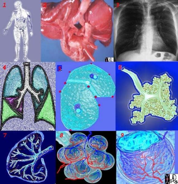
Biological Unit
In the world of the lung a reasonable starting structural and functional unit is the alveolus. The tubular airways are the specialized conduits for one of the the links between the alveolus with its surrounding environment. As in all instances in biology the alveolus is made of smaller parts which for the sake of simplicity will be discussed further as the module advances. The alveolus joins with other alveoli to form the lobule, and the lobules join to form segments, which in turn form lobes which combine to form the lungs themselves. The respiratory bronchioles join to form the terminal bronchioles, which in turn join to form bronchioles proper, which subsequently become segmental bronchi, lobar bronchi, mainstem bronchi and then the trachea. The traches communicates with the outside world via the oropharynx. The lungs are protected and supported by a double layer of outer capsule called the pleura, with the rib cage, associated muscles, and skin representing additional layers of support and protection.
42651c
keywords lung chest Ashley Davidoff TheCommonVein.net
Structure
Structure and function of the lung work hand in hand. The order of one is dependent on the order of the other and similarly when there is disorder of one there is inevitably disorder of the other. In the study of structure and function we tend to separate the two in an attempt to understand each of them, but of course in biology they are inextricably linked and dependent on one another. Within the realm of the biological units, common structural features and functional features exist, whether we are observing the cell , the tissue, the organ, the body or the larger communities.
From a structural point of view the lungs are large, voluminous, pyramidal in shape and together form a bell shaped organ. They are found in the chest cavity, one on the right side and one on the left. They are spongy in nature.
Parts
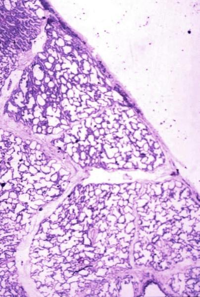
Normal lung histology
This image is a panoramic view of the lung showing secondary lobules and interlobular septa. Within the interalveolar septae, one sees small venules and lymphatics.Courtesy Armando Fraire MD. 32649b
code lung pulmonary alveoli alveolus secondary lobule interlobular septa vein lymphatic histology
interstitium interstitial
Courtesy of: Armando Fraire, M.D. Ashley Davidoff TheCommonVein.net
32649b
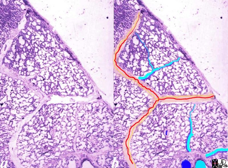
Normal lung histology
This image is a panoramic view of the lung showing secondary lobules and interlobular septa. Within the interalveolar septae, one sees small venules and lymphatics .
The side by side images show the interlobular septa within which reside the pumonary venules (red) and lymphatics and within the center of the lobule run the respiratory bronchioles (teal) and pulmonary arterioles (blue)
Courtesy Armando Fraire MD. 32649b
key words
lung pulmonary alveoli alveolus secondary lobule interlobular septa vein lymphatic histology
interstitium interstitial
Courtesy of: Armando Fraire, M.D. Ashley Davidoff TheCommonVein.net
32649c06.8s
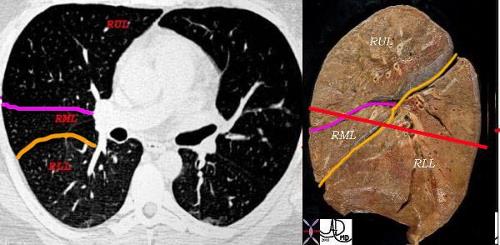
This combination shows the two fissures of the right lobe on the CT scan separating the RLL from the RML (major fissure in orange) and the RML from the RUL. (minor or transverse fissure in pink) The anatomic specimen is in the sagittal plane with the posterior aspect to your right. The red line drawn through the specimen represents the level of the X-sectional plane revealing in sequence from posterior to anterior, the RLL, major fissure, RML, minor fissure, RUL. On the left side there is the faint hint of hypovascularity along the major fissure.
Ashley Davidoff MD. TheCommonVein.net 32160c2lb_6
The parts of the lungs can be subdivided in one of many ways but in the end there are the substantive components, and the connecting links that link the parts to each other and the parts to a bigger whole.
So for example the lung cells are held together by cellular bonds and connective tissue, and are linked to each other and the other organs by air ducts blood vessels nerves and lymphatics. These components at the microscopic level unite and form a lung lobule, which in turn unite in additive fashion to form lobes and finally the organ itself.
Size
key words chest lung CXR normal imaging radiology
Ashley Davidoff MD TheCommonVein.net 41818b
The chest quietly expands and contracts under basal conditions in order to serve the alveoli. At first glance it seems like a simple bellows-like process, but as one delves into the layers of detail, the complexity of the structural design unfolds as a combination of physical and chemical forces.
Ashley Davidoff MD TheCommonVein.net 42530b05b09b28
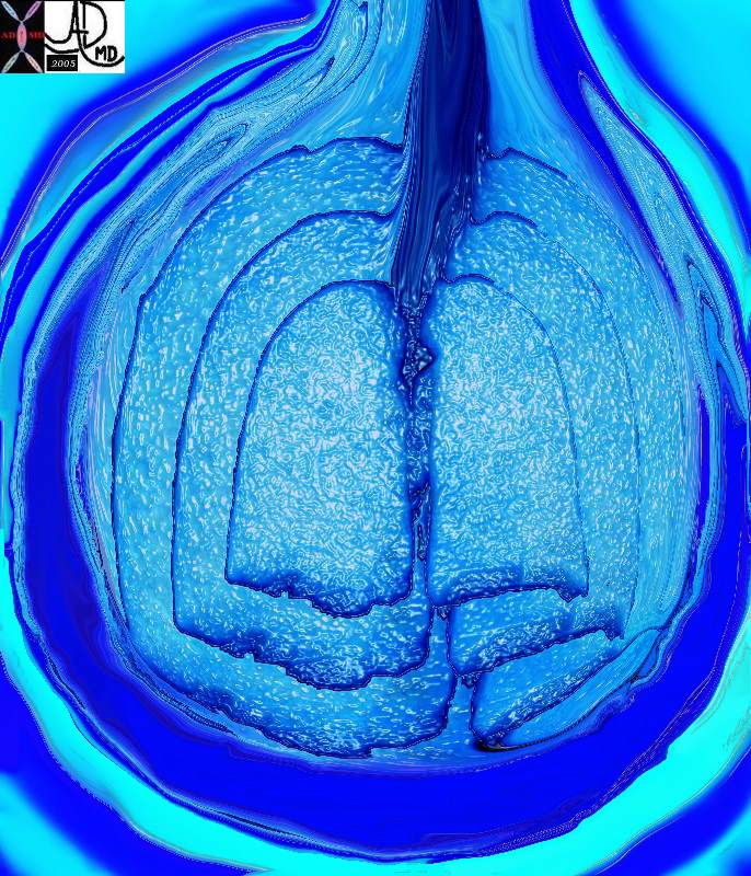
As described shape can reflect a changing size because a sheer increase in volume or pressure in a structure will be reflected in the shape. The are many other factors that affect shape often caused by forces within the structure or the forces that surround the structure.
There are many references to the shape bell in biology architecture and statistics. The thoracic cage in this instance is bell shaped.
Ashley Davidoff MD TheCommonVein.net . 61829
As described shape can reflect a changing size because a sheer increase in volume or pressure in a structure will be reflected in the shape. The are many other factors that affect shape often caused by forces within the structure or the forces that surround the structure.
Position
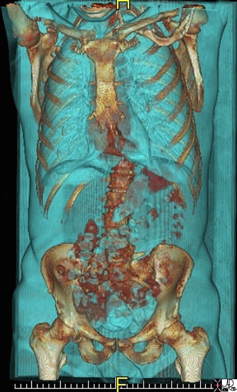
keywords abdomen chest space cavity cavities normal anatomy structure CTscan 3D
Ashley Davidoff MD TheCommonVein.net 71198.800
Character
Character is often the most difficult of the structural principles to grasp. In our inquisitive way we “taunt” structure by subjecting it to any one of the forces in our armamentarium and seeing how it reacts. We pull or push on it, throw different sorts of temperatures, chemical, radiational or magnetic fields toward it, and we observe its reaction. The reaction of the lung tissue to the force we subject it to is labelled its character. If one pulls the two ends of an elastic band apart and thereafter release them – they will return to their original position. It is thus characterized as having elastic properties. At the cellular level we stain the cells with different chemicals and observe how they react, at the clinical level we palpate and percuss, while at the imaging level we subject the tissues to ultrasound waves, X-rays, and changing magnetic forces in order to characterize them.
Normal Character on H and E Stain
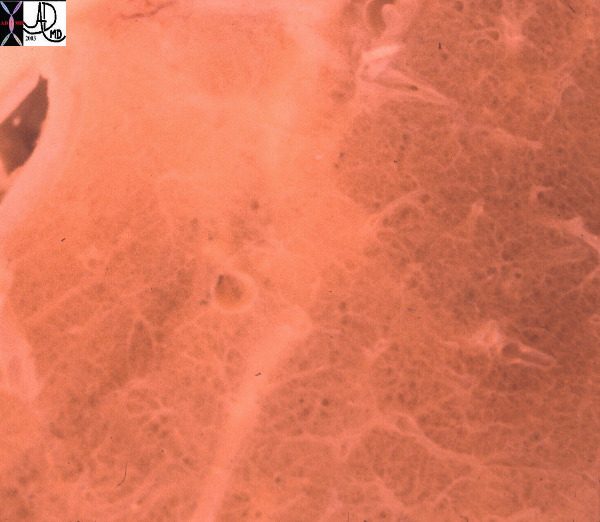
Normal lung histology
Courtesy of: Armando Fraire, M.D. Ashley Davidoff TheCommonVein.net
32649b

Lower magnification of the lung with H and E stain shows cup-shaped alveolar spaces outlined by delicate thin alveolar capillary membrane.
key words
lung, pulmonary, normal alveolus, alveoli, histology, interstitium, interstitial
Courtesy Armando Fraire MD. 32819
Normal Appearance on Gross Anatomy – Spongy
The lungs have a spongy feel macroscopically characterised by small air filled holes
Ashley Davidoff MD TheCommonVein.net 32300d
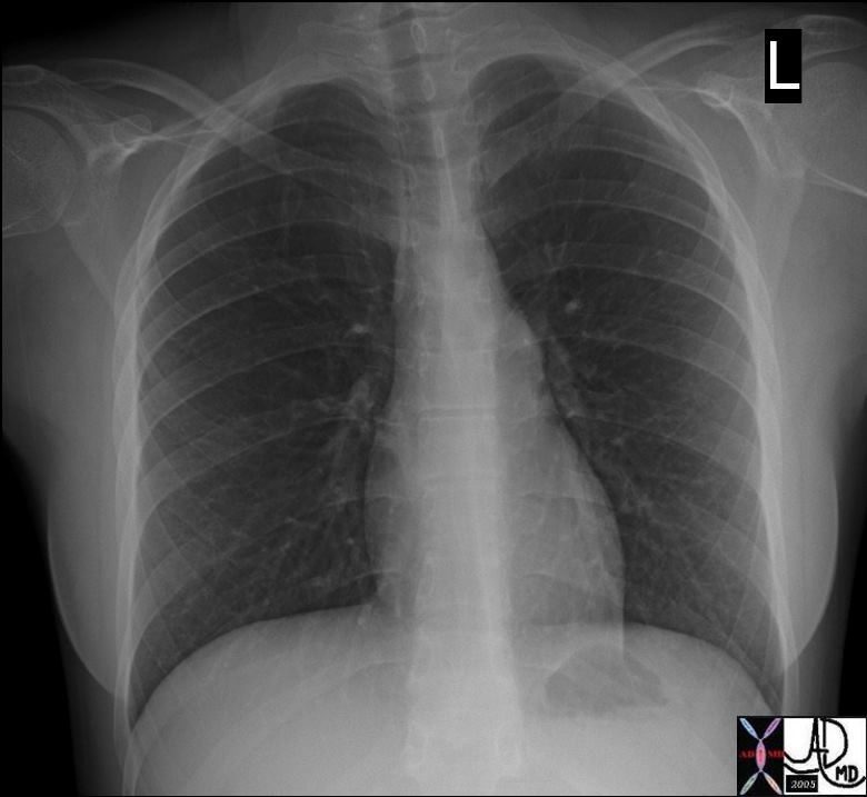
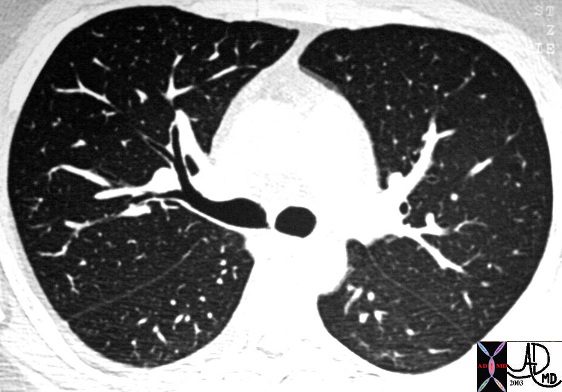
Normal Characteristics on X-Ray
key words chest lung CXR normal imaging radiology
Ashley Davidoff MD TheCommonVein.net 41818b
Ashley Davidoff MD TheCommonVein.net 30524
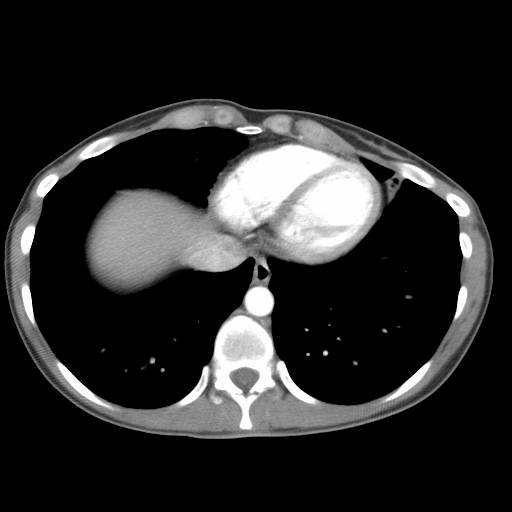
This is a normal CT examination of the chest through the level of the heart with contrast showing the densities of the lung, heart, contrast, soft tissues and bones on soft tissue windows
Ashley Davidoff MD TheCommonVein.net 33755
Links Connections
The lungs on the one end are connected to the outside environment, connected to each other via the airways and connected to the rest of the body via both the systemic and the pulmonary circulation of arteries, veins, and lymphatics. It has intimate connection with the rest of the nervous system through the sympathetic and parasympathetic nervous system, while connective tissue, ligaments, mesenteries and cement matrix bond and link its tissues.
Time Growth and Aging
The universal element of time applies through all aspects of biology, whether one is talking about the lifespan of the a red cell or whether one is talking about the growth and development of the embryo to adulthood. Time and cycles are inextricably linked to biology.
Units to Unity
Uniqueness
The structure and function of the lung is absolutely essential and unique. From a structural point of view for example there is no other structure that has its spongy feel, and no other structure can perform its delivery and exchange of gases in the way that it does. The kidney is the other large filter in the body, but it is specialized to filter fluids rather than gases.. As each cell is studied in depth, morphological uniqueness will be appreciated in the same way that each human being is recognized as unique.
Shape is well understood by us all but more subjective under many circumstances. In the case of a circle or a sphere we can all imagine and appreciate the perfect sphere. In the biological world perfect spheres or cuboids rarely exist . We use the word cuboidal for example to transmit the idea of a cube like structure. Either way we can communicate shape imagine All structures are also made up of parts.
Dependance and Independence
The lung cannot function alone. On the one end it needs the outside environment for its raw material, and its vascular connections to deliver its products. It needs the nervous system to control its function, the endocrine system and nutritional system of the body to maintain its metabolism and viability, and the filtering function of the kidneys to keep a clean environment. The bony rib cage and associated muscular systems are vital to help move the air back and forward. It needs the macrophages and the reticuloendothelial system to keep the environment protected.
Time – Embryology growth and Aging
Space
Space is a second element that is inextricably linked to biology. All biological units need space to live and to breathe and to work in. Encroachment on another’s space is a physical and moral transgression.
Forces
In addition to space and time, there are other forces in our environment that cause the environment to continually change. There are some forces like gravity that are always present on earth and do not change while others like temperature, and humidity are in continual flux. These forces which also include chemical , radiational , and mechanical forces create an ongoing minute to minute challenge for the organism.
Interactions
Biological units interact with the forces of the environment resulting in a change. The exchange may lead to more order or more disorder.
States of Being
There are states of order and states of disorder. In the extreme we have life and death, and in between our lives exist between states of relative order and states of relative disorder.
Health and Disease
In general the states of order are called health and the states of disorder are called disease.
The Field of Medicine
The field of medicine focuses on the states of order and disorder. The basic elements that allow the physician to accomplish part of the mission is the understanding of structure, function and disease, and to responsibly and wisely diagnose and treat disorder.
The goal of this work is to provide a path for the medical student who comes to us on the first day of his/her education and says – :”I know nothing and I want to grow into a compassionate, thoughtful, knowledgeable, and wise physician.”
It is also for those who want to understand how the world within us and around us have common universal organization and principles