
Findings of a right upper lobe collapse (atelectasis) typically present characteristic signs due to the volume loss in the affected lobe and the secondary changes in surrounding structures. Some of the common findings include:
Ashley Davidoff MD TheCommonVein.net
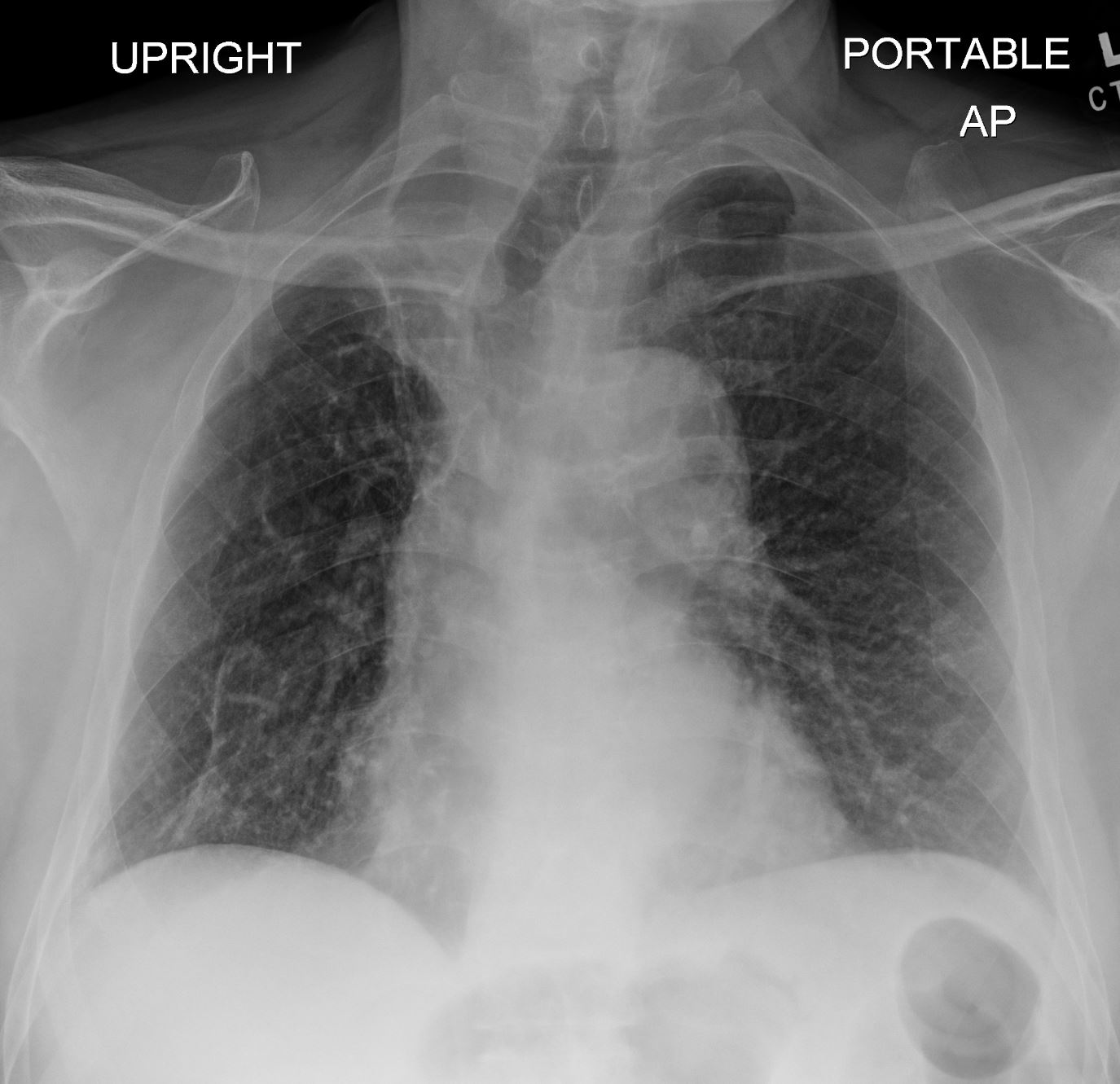
This combination of images shows the manifestations of a malignant mass in the hilum causing compression of the right mainstem bronchus. The PA CXR shows signs of volume loss (atelectasis characterized by elevation of the right hemidiaphragm (black arrowhead), rightward tracheal and mediastinal shift (white arrowheads) and elevation of the minor fissure contributing to the reverse S sign of Golden. There is a vague infiltrate in the right upper lobe correlating with an anterior pie shaped density of the lateral (blue arrowheads), consistent with collapse of the anterior segment of the RUL
32292cL01
Ashley Davidoff MD TheCommonVein.net
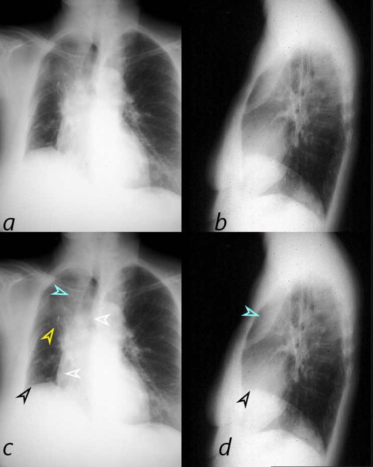
The scout film performed prior to a CT scan from a 76 year old man with chest pain and shortness of breath. The appearance suggests atelectasis of the right upper lobe with the normal position of the minor fissure (yellow) altered so that the upper portion (light green above the yellow line) is shifted upward caused by volume loss of atelectatic right upper lobe. (pink). The lower portion of the fissure (light green below the yellow line) is bulging rightward and outward caused by an implied mass (dark green). The “reversed S sign of Golden” is demonstrated in this case and infers a central mass causing obstruction and resulting in the shape described by the light green line of the minor fissure.
Ashley Davidoff
TheCommonVein.net
87740c02.8s
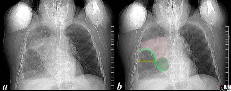
59year old male presents with cough. Chest Xray reveals signs of segmental right upper lobe atelectasis with elevation of the minor fissure and right hemidiaphragm confirms the presence of atelectasis and an air bronchogram. A proximal obstructing lesion was suspected. Slightly enlarged lymph nodes are noted in the azygous region (last row)
Ashley Davidoff MD TheCommonVein.net
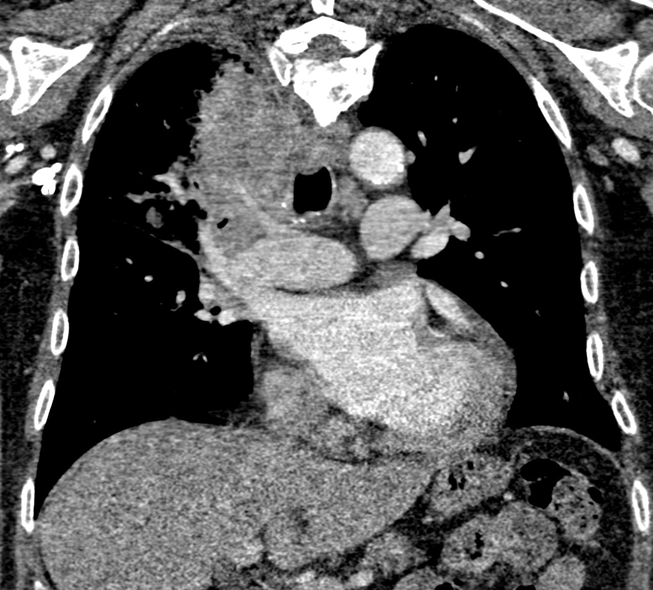
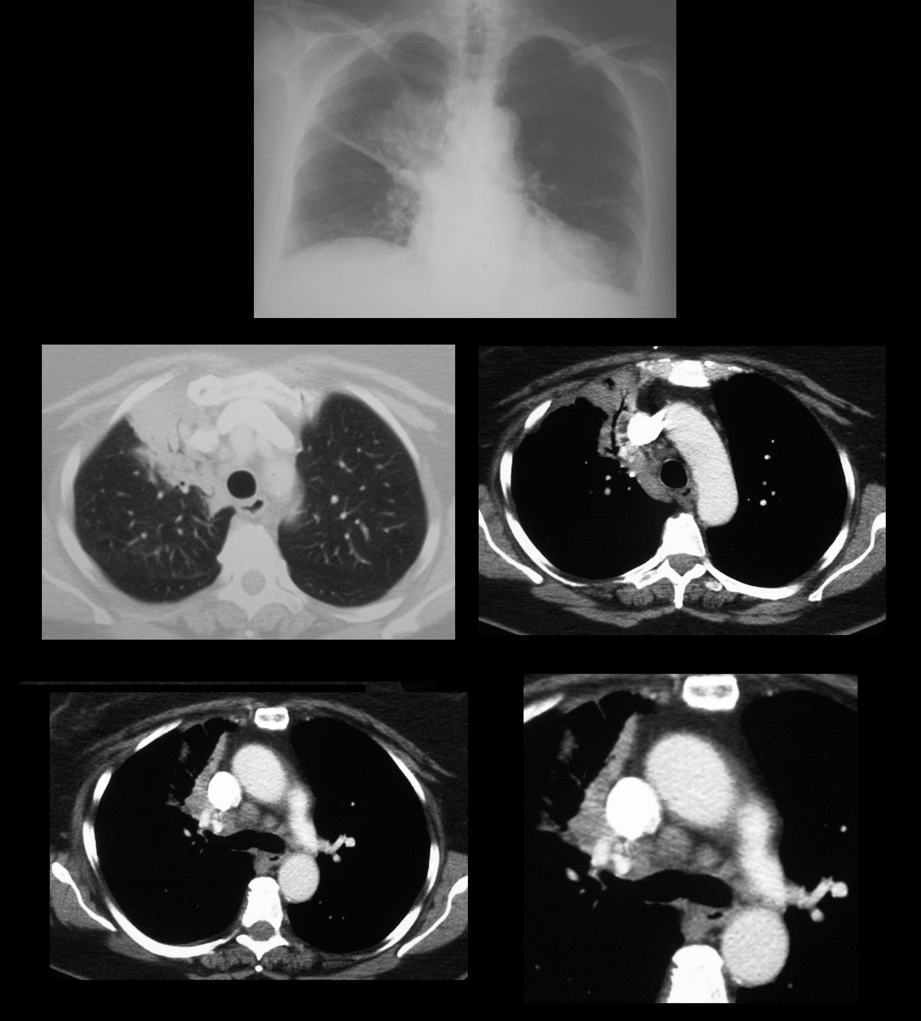
There is patchy opacity in the right upper lung
which may represent post obstructive atelectasis from the right hilar mass or multifocal airspace disease. There is encasement of the right upper lobe pulmonary artery suggesting a malignant process.
Ashley Davidoff MD
TheCommonVeiin.net
70M lung ca 005
- Volume Loss and Collapse of the Right Upper Lobe:
- The right upper lobe appears shrunken or collapsed.
- The collapsed lobe may present as a triangular or wedge-shaped opacity, particularly visible on cross-sectional imaging.
- The interlobar fissure (minor and major) may be displaced toward the right upper lobe due to volume loss.
- Elevation of the Right Horizontal Fissure:
- The minor (horizontal) fissure is typically displaced upward due to the loss of volume in the right upper lobe.
- The major fissure may also move superiorly to fill the space left by the collapsed lobe.
- Mediastinal Shift:
- There may be a shift of the mediastinum (heart, trachea, and great vessels) toward the right (the side of the collapse) as the lung volume decreases, especially in more severe cases.
- Crowding of Bronchovascular Structures:
- The bronchi and vessels within the right upper lobe may appear crowded or distorted due to the collapse.
- Hilum Shift:
- The right hilum, where the bronchi and vessels enter the lung, may also shift superiorly as the upper lobe contracts.
- Compensatory Hyperinflation of Adjacent Lobes:
- The right middle and lower lobes may appear hyperinflated or overexpanded to compensate for the lost volume of the collapsed upper lobe.
- Golden S Sign (in cases of obstructing mass):
- In some cases where the collapse is due to a central mass (such as a tumor), the Golden S sign may be seen. This sign refers to an S-shaped curvature of the minor fissure caused by the mass pushing the fissure upward, while the collapsed lobe pulls it inward.
- Opacity in the Affected Lobe:
- The collapsed upper lobe may appear as a dense opacity on imaging, typically located in the upper portion of the right lung.
- Air Bronchograms:
- If the collapse is non-obstructive, air bronchograms (air-filled bronchi seen within the collapsed, opaque lung) may be visible, helping to differentiate the collapse from other conditions like consolidation.
- Associated Pleural Changes:
- Chronic collapse may be associated with pleural thickening or even mild pleural effusion.