Total lung collapse refers to the complete atelectasis of an entire lung, resulting in loss of aeration and significant volume reduction. It is caused by a variety of conditions, most commonly central airway obstruction or external compression. Importantly, tension pneumothorax can also result in total lung collapse due to the accumulation of air in the pleural space under pressure, compressing the lung and shifting mediastinal structures.
Radiological Features
- Chest X-Ray (CXR):
- Complete Opacification:
- The affected hemithorax appears completely opaque (obstructive or compressive collapse) or hyperlucent (tension pneumothorax).
- Mediastinal Shift:
- Toward the affected side: Seen in obstructive collapse.
- Away from the affected side: Seen in tension pneumothorax.
- Elevation of the Hemidiaphragm:
- The diaphragm on the affected side is elevated in obstructive or compressive collapse.
- Crowding of Ribs:
- Narrow intercostal spaces on the side of obstructive collapse.
- Widened intercostal spaces in tension pneumothorax due to pleural pressure.
- Absence of Lung Markings:
- In pneumothorax, there is no vascular lung pattern distal to the collapsed lung.
- Complete Opacification:
- CT (High-Resolution CT):
- Lung Collapse:
- The lung appears as a dense, shrunken structure adjacent to the mediastinum or pleura.
- Airway Obstruction:
- Identification of the causative lesion in obstructive collapse (e.g., tumor, mucus plug, foreign body).
- Tension Pneumothorax:
- Hyperlucency in the pleural space with compression of the collapsed lung.
- Mediastinal Shift Away from the Collapsed Lung: A hallmark feature.
- Flattening or inversion of the ipsilateral hemidiaphragm.
- Associated Features:
- Pleural effusion or air-fluid levels (e.g., hydropneumothorax).
- Lung Collapse:
Causes of Total Lung Collapse
- Obstructive:
- Endobronchial Tumors:
- Primary lung cancer or metastatic disease.
- Foreign Body:
- Often in children.
- Mucus Plug:
- Common post-operatively or in conditions like asthma or cystic fibrosis.
- Endobronchial Tumors:
- Compression:
- Pleural Effusion:
- Large effusions leading to compressive atelectasis.
- Mass Effect:
- Mediastinal or chest wall tumors compressing the lung.
- Pleural Effusion:
- Tension Pneumothorax:
- Accumulation of air under pressure in the pleural space.
- Causes complete lung collapse by compressing the lung tissue.
- Key Differentiating Features:
- Mediastinal shift away from the affected side.
- Severe flattening or inversion of the diaphragm.
- Widened intercostal spaces on the affected side.
- Trauma:
- Rib fractures with lung puncture causing pneumothorax or hemothorax.
Differential Diagnosis
- Large Pleural Effusion:
- Uniform opacification but with a clear fluid level on upright imaging.
- Complete Pneumonia:
- Diffuse consolidation without volume loss or mediastinal shift.
- Pulmonary Fibrosis:
- Chronic condition with associated architectural distortion.
Complications
- Tension Pneumothorax:
- Life-threatening condition with hemodynamic compromise due to decreased venous return and cardiac output.
- Requires immediate recognition and decompression.
- Hypoxemia:
- Due to significant ventilation-perfusion mismatch.
- Secondary Infections:
- Increased risk of pneumonia or abscess formation.
Management
- Tension Pneumothorax:
- Immediate needle decompression or chest tube placement.
- Obstructive Collapse:
- Bronchoscopy to relieve obstruction (e.g., foreign body, mucus plug).
- Biopsy for suspected malignancy.
- Compression Collapse:
- Thoracentesis for pleural effusion.
- Chest tube insertion for pneumothorax or hemothorax.
Clinical Relevance
Including tension pneumothorax as a cause of total lung collapse highlights the importance of recognizing this life-threatening condition. Its radiological features, such as hyperlucency, mediastinal shift away from the collapsed lung, and widened intercostal spaces, make it distinct from obstructive and compressive causes. Early intervention is crucial to prevent fatal outcomes.
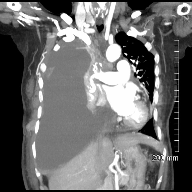
85-year-old female with a history of lung cancer, presents with a dyspnea and hypotension. CT scan shows a large right pleural effusion under pressure, with mediastinal shift to the left. In addition, there is compression of the heart with back up of venous return due the pressure effect on the heart and vascular structures. The effusion in the right pleural cavity with atelectatic lung herniates into the left hemithorax.
Ashley Davidoff MD TheCommonVein.net 106Lu 118467
85-year-old female with a history of lung cancer, presents with a dyspnea and hypotension. CT scan shows a large right pleural effusion under pressure, with mediastinal shift to the right. In addition, there is compression of the heart with back up of venous return due the pressure effect on the heart and vascular structures. Among the structures showing venous distension are the SVC (blue arrowhead, a) right sided upper limb veins (blue arrowhead b) and the left upper pulmonary veins (red arrowhead, b. The effusion in the right pleural cavity with atelectatic lung herniates into the left hemithorax, (white arrowhead, c). There is a dense sediment in the pleural fluid (red arrowhead, d) suggesting blood in the pleural cavity. The left atrium is compressed (maroon arrowhead, d)
Ashley Davidoff MD TheCommonVein.net106Lu 118467c

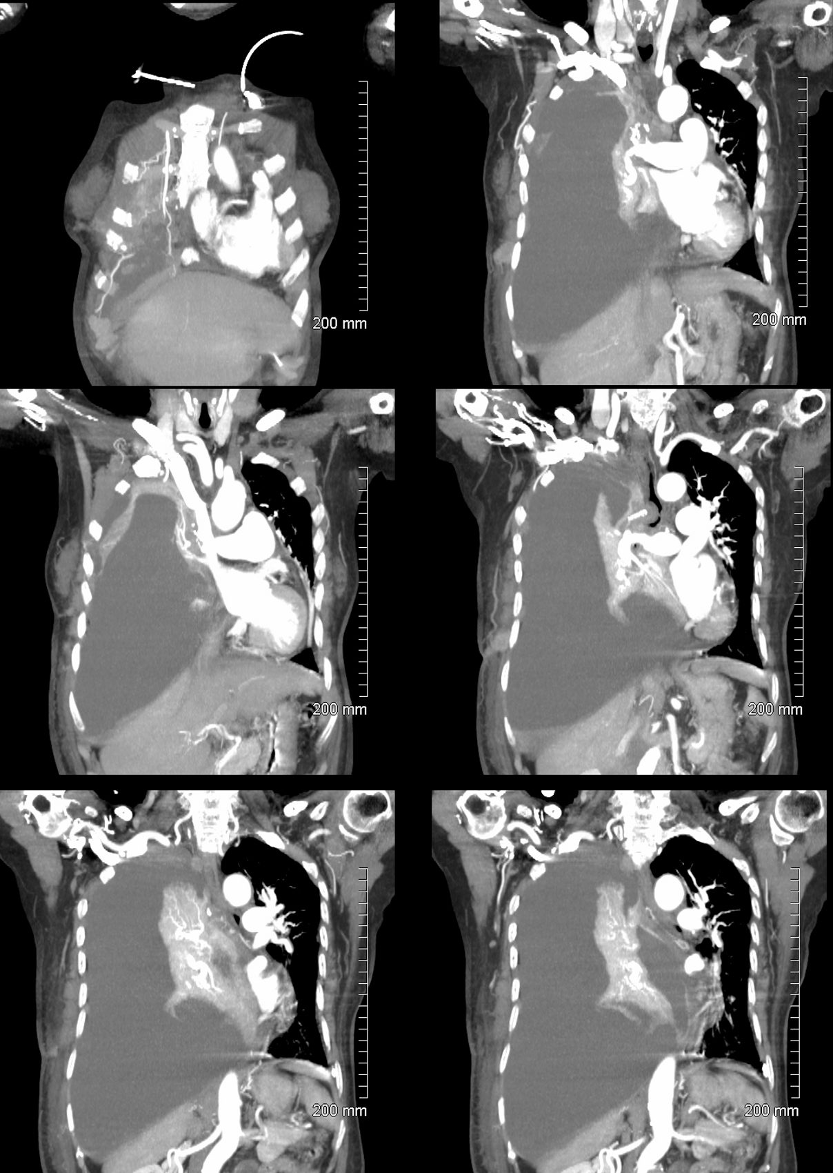
85-year-old female with a history of lung cancer, presents with a dyspnea and hypotension. Reconstruction of the CT scan in the coronal plane, shows a large right pleural effusion under pressure with herniation into the left chest (white asterisk e,and f) , with mediastinal shift to the left (yellow arrowhead b, c, d). In addition, there is compression of the heart with back up of venous return due the pressure effect on the heart and vascular structures. Among the structures showing venous distension are the SVC (blue arrowhead, c) right sided upper limb veins (blue arrowhead d) and the left upper pulmonary veins (red arrowhead, d and f). The density of the systemic venous and arterial systems is similar, but vascular structures as noted by the green arrowhead in a could represent venous collaterals.
Ashley Davidoff MD TheCommonVein.ne 106Lu 118467cL
Radiologic Findings of Total Lung Collapse
- Increased Opacity:
- The collapsed lung becomes densely opaque on imaging, as the air is absorbed from the lung tissue and the remaining lung is composed primarily of soft tissue.
- The entire hemithorax (one side of the chest) may appear more opaque than the unaffected side.
- Mediastinal Shift:
- Structures such as the heart, trachea, and mediastinum shift toward the collapsed lung due to the loss of volume on that side.
- This shift can be prominent, especially in cases of complete lung atelectasis without compensatory hyperinflation on the contralateral side.
- Elevation of the Hemidiaphragm:
- The diaphragm on the side of the collapsed lung is pulled upward due to the decreased intrathoracic pressure on that side.
- The hemidiaphragm may be noticeably higher than the one on the unaffected side.
- Rib Space Narrowing:
- There is narrowing of the intercostal spaces on the side of the collapse, as the chest wall is pulled inward by the loss of lung volume.
- Silhouetting of Adjacent Structures:
- The normal borders of structures like the heart, aorta, and hemidiaphragm may be obscured by the collapsed lung, which now occupies a smaller, dense space adjacent to these structures.
- This finding can help in determining the location of the collapse and distinguishing it from other causes of increased opacity.
- Compensatory Hyperinflation of the Opposite Lung:
- The unaffected lung may appear hyperinflated, as it expands to fill some of the space left by the collapsed lung.
- The expansion of the unaffected lung may include herniation across the midline into the space of the collapsed lung.
- Crowding of Pulmonary Vessels and Bronchial Structures:
- Bronchi and vessels within the collapsed lung appear crowded and may be displaced toward the hilum.
- Bronchial Cutoff Sign (If Visible):
- On CT or sometimes X-ray, the cause of the collapse, such as an obstructing tumor or mucus plug, may be visible as an abrupt cutoff in the bronchus.
Additional Findings on CT
CT imaging provides a clearer view and may show:
- Direct visualization of the obstruction if there is a mass, mucus plug, or foreign body.
- Collapsed lung tissue appearing as a dense, triangular or wedge-shaped opacity.
- Possible pleural effusion on the affected side, sometimes seen in cases of malignancy or infection.
Causes
Common causes of total lung collapse include:
- Endobronchial obstruction: Often due to a tumor, mucus plug, or foreign body in the main bronchus.
- Malignancy: Lung cancer can obstruct a major bronchus and is a common cause of total lung collapse in adults.
- Large pleural effusion or pneumothorax: Can indirectly cause lung collapse by compressing the lung from outside.
Summary
Total lung collapse results in a range of radiologic findings, primarily marked by increased opacity, mediastinal shift, elevation of the hemidiaphragm, narrowing of the rib spaces, and potential compensatory hyperinflation of the opposite lung. These findings are important to identify the presence and side of the collapse and suggest possible underlying causes that may require further evaluation.
Total Left Lung Collapse
Tension Pneumothorax
Ashley Davidoff MD TheCommonVein.net
77949
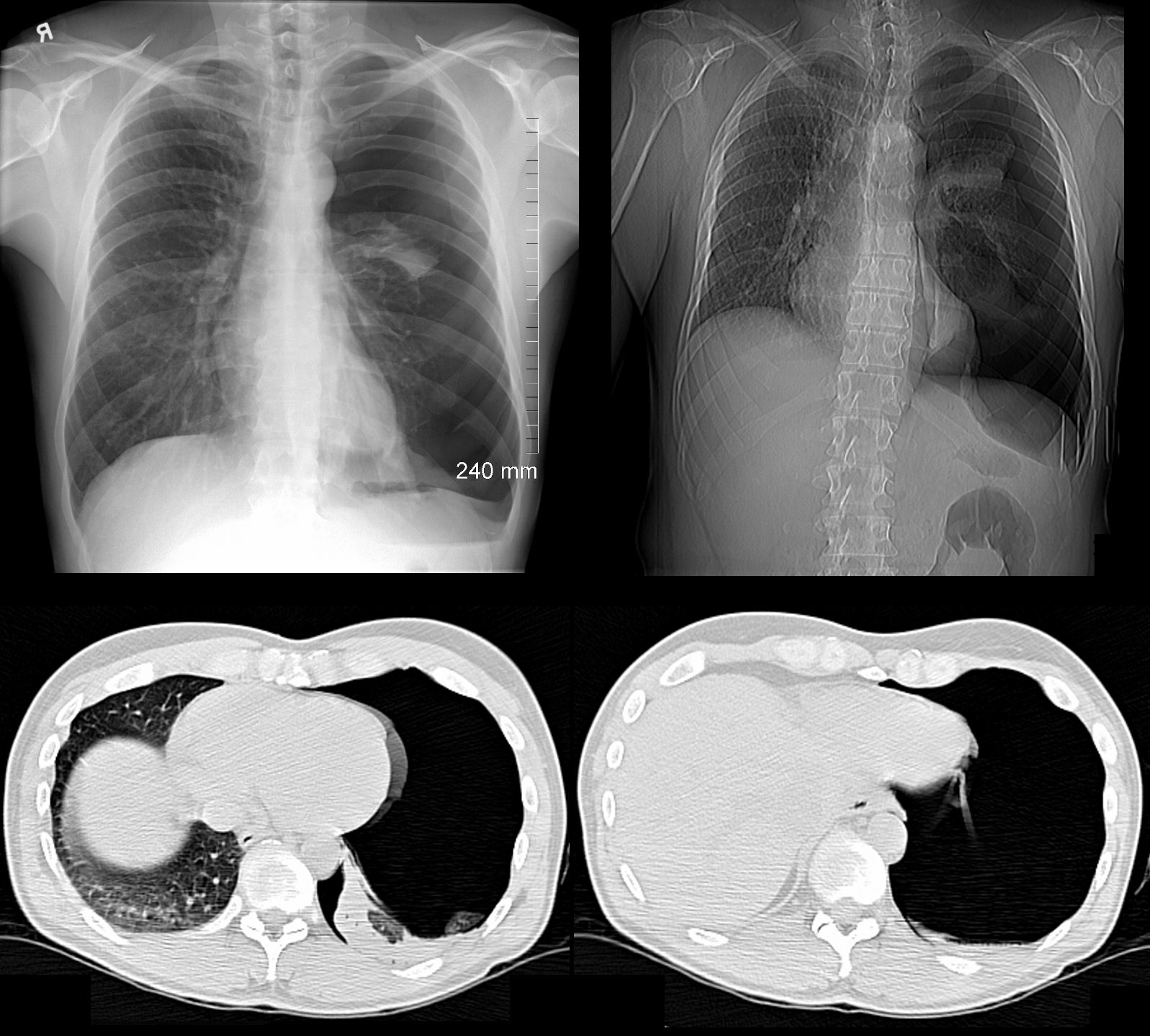
49 year old male with a cough presents for a Chest Xray which showed a tension pneumothorax. Chest tube was placed emergently in the radiology department.
Ashley Davidoff MD TheCommonVein.net
117300c
Total Right Lung Collapse
Tension Hydrothorax
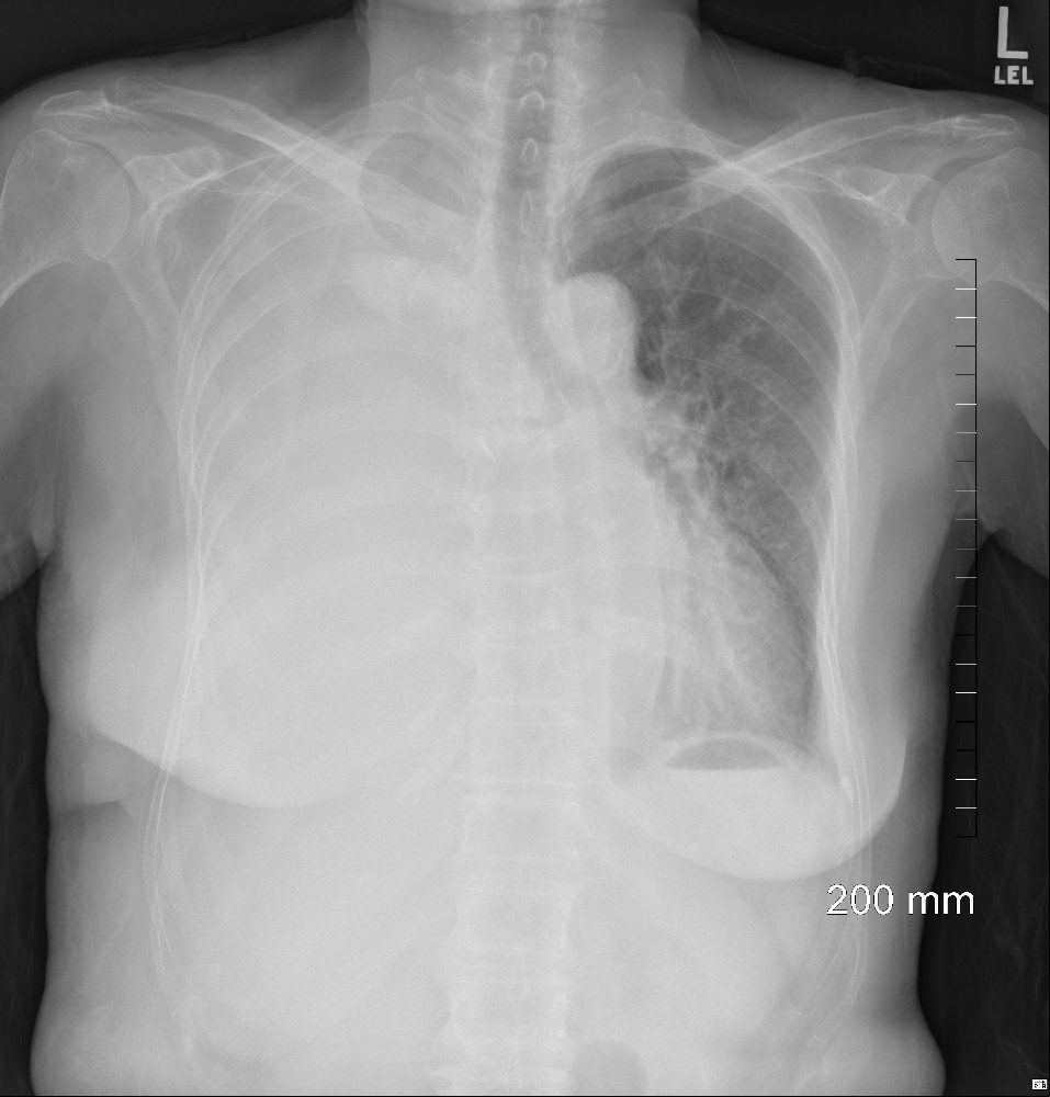
from large pleural effusion and probable hemothorax under tension with atelectasis of the right lung
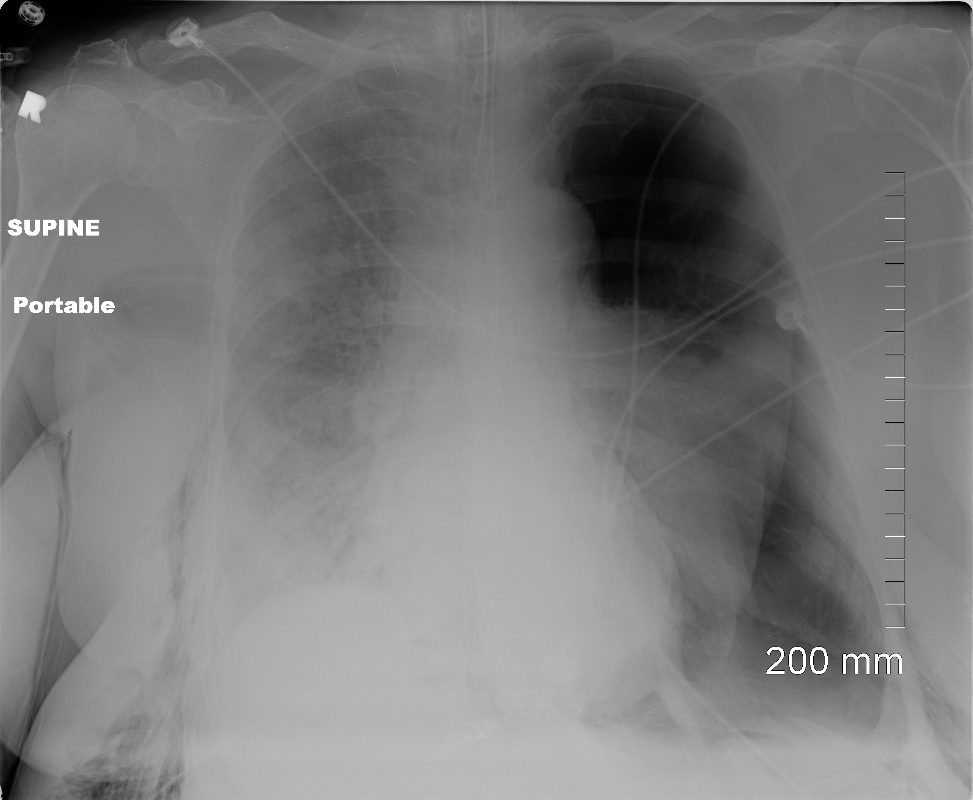
85-year-old female with a history of lung cancer, presents with dyspnea and hypotension. CXR shows white out of the right hemithorax with pressure effect characterised by narrowing of the distal trachea cardio-mediastinal shift to the left and atelectasis in the left lower lobe.
Ashley Davidoff MD TheCommonVein.netSee 106Lu 118468
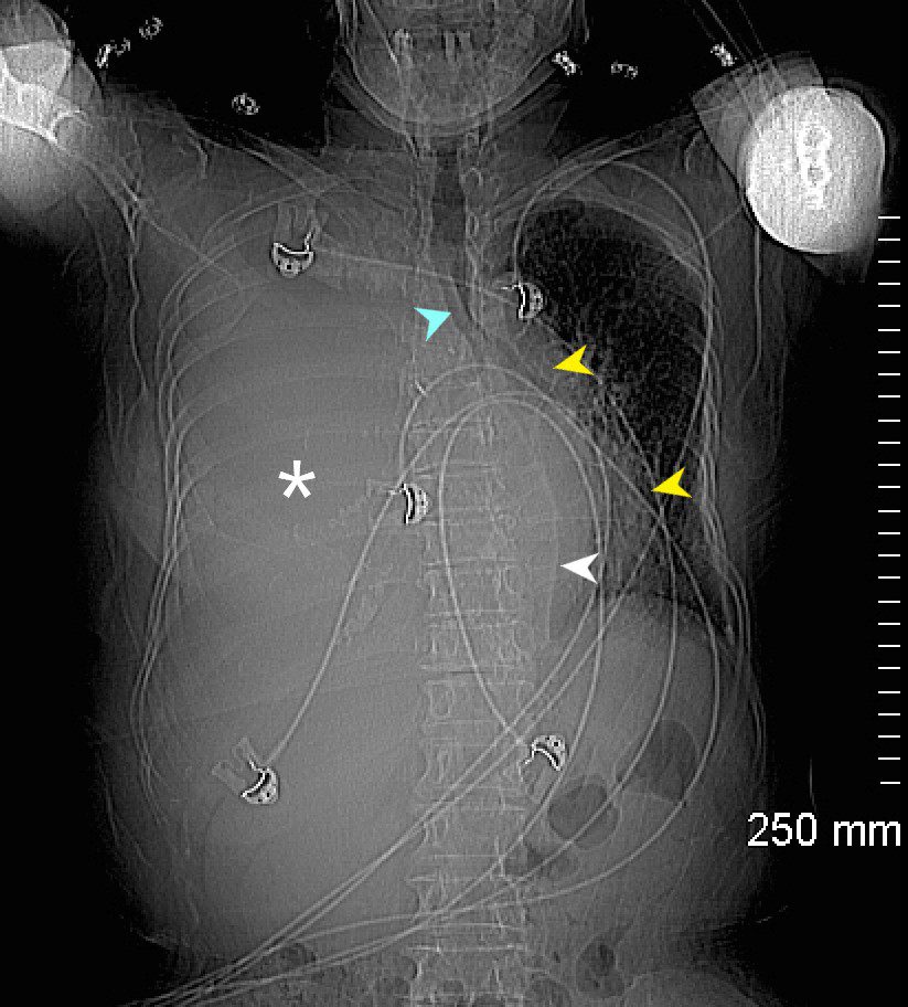
85-year-old female with a history of lung cancer, presents with a dyspnea and hypotension. Scout film prior to the CT scan shows “white out” of the right hemithorax (white asterisk) with pressure effect characterised by narrowing of the trachea (blue arrow) mediastinal shift yellow arrows) and herniation into the left chest characterised by a leftward shift of the azygo-esophageal junction line (white arrow).
Ashley Davidoff MD TheCommonVein.ne 106Lu 118463L

85-year-old female with a history of lung cancer, presents with a dyspnea and hypotension. CT scan shows a large right pleural effusion under pressure, with mediastinal shift to the right. In addition, there is compression of the heart with back up of venous return due the pressure effect on the heart and vascular structures. Among the structures showing venous distension are the SVC (blue arrowhead, a) right sided upper limb veins (blue arrowhead b) and the left upper pulmonary veins (red arrowhead, b. The effusion in the right pleural cavity with atelectatic lung herniates into the left hemithorax, (white arrowhead, c). There is a dense sediment in the pleural fluid (red arrowhead, d) suggesting blood in the pleural cavity. The left atrium is compressed (maroon arrowhead, d)
Ashley Davidoff MD TheCommonVein.net106Lu 118467c

85-year-old female with a history of lung cancer, presents with a dyspnea and hypotension. Reconstruction of the CT scan in the coronal plane, shows a large right pleural effusion under pressure with herniation into the left chest (white asterisk e,and f) , with mediastinal shift to the left (yellow arrowhead b, c, d). In addition, there is compression of the heart with back up of venous return due the pressure effect on the heart and vascular structures. Among the structures showing venous distension are the SVC (blue arrowhead, c) right sided upper limb veins (blue arrowhead d) and the left upper pulmonary veins (red arrowhead, d and f). The density of the systemic venous abd arterial systems is similar, but vascular structures as noted by the green arrowhead in a could represent venous collaterals.
Ashley Davidoff MD TheCommonVein.ne 106Lu 118467cL

85-year-old female with a history of lung cancer, presents with a dyspnea and hypotension. CT scan shows a large right pleural effusion under pressure, with mediastinal shift to the left. In addition, there is compression of the heart with back up of venous return due the pressure effect on the heart and vascular structures. The effusion in the right pleural cavity with atelectatic lung herniates into the left hemithorax.
Ashley Davidoff MD TheCommonVein.net 106Lu 118467
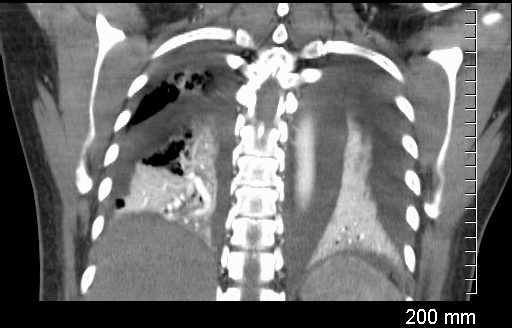
Coronal CT through the lungs show bilateral pleural effusions with compressive atelectasis
Ashley Davidoff MD TheCommonvein.net
238Lu
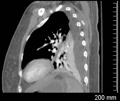
Sagittal CT through the left lung shows undulations of the posterior surface of the left lung, and the suggesting differing pressures on the lung parenchyma by the effusions and indicating complexity and loculation.
Ashley Davidoff MD TheCommonVein.net
238Lu
Whole Lung Atelectasis Due to Obstruction
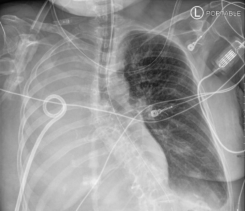
59F shows total white out caused by collapse of right lung with an
occluded right main step bronchus associated with a
large right sided effusion. The occlusion is likely due to proximal cancer. A pigtail drain has been placed to drain the effusion
Ashley Davidoff MD TheCommonVein.net 104 Lu
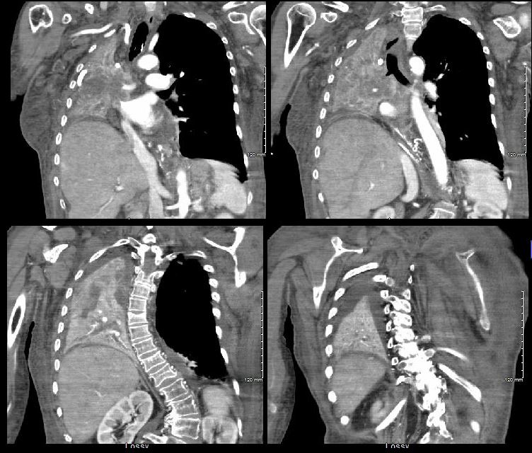
59F shows total collapse of left lung with an
occluded right main step bronchus(top right image)associated with a
right sided effusion. The occlusion is likely due to proximal cancer
Ashley Davidoff MD TheCommonVein.net 104 Lu
Total Lung Compressive Atelectasis
Effusion
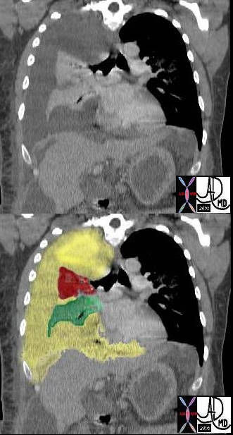
In this case there a large right sided pleural effusion (yellow) with secondary atelectasis of the right lung. (red and green) This coronal CT of the chest at the level of the left ventricle shows a large right pleural effusion which lies between the visceral and parietal pleura. Once the effusion is large enough to weaken the capillary forces that hold the parietal and visceral pleura together, it fail, and the lung collapses which is what is noted on this image – ie total lung collapse because of loss of cohesive adhesive forces.
Courtesy of: Ashley Davidoff, M.D. TheCommonvein.net 42558c
White Out of the CXR with Passive Compressive Atelectasis of the Left Lung
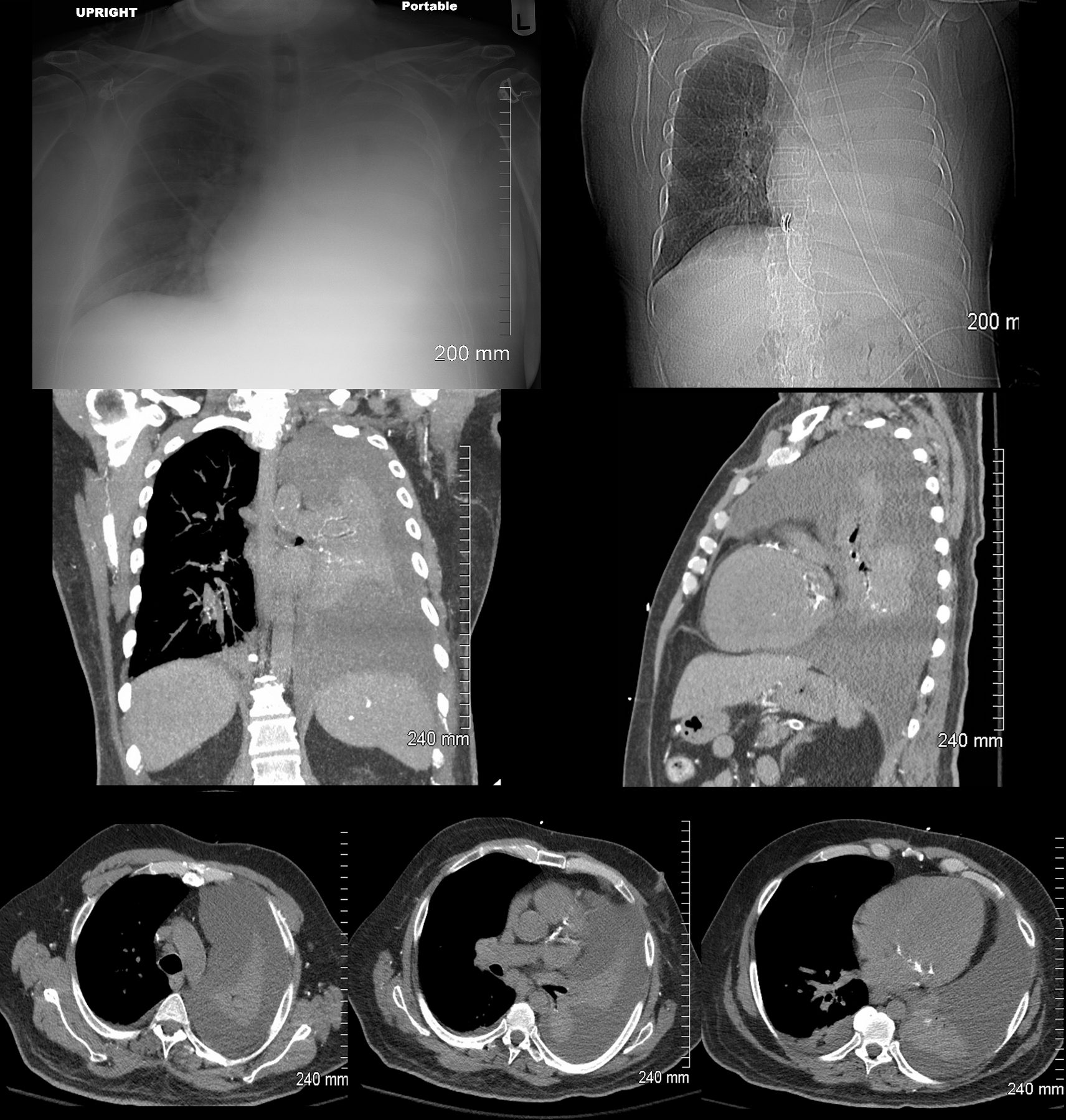 White Out of the CXR with Passive Compressive Atelectasis of the Left Lung
48 year-old male presents with a dyspnea. CXR shows a total white out of the left chest with pulmonary congestion. CT scan shows a large left pleural effusion with total atelectasis of the left lung. Incidental note is made of premature calcific coronary artery disease.
Ashley Davidoff MD TheCommonVein.net
Links and References
Chat GPT Radiological findings of total lung collapse aka total lung atelectasis
