
Wegener’s Granulomatosis GPA Granulomatosis with Polyangiitis
81-year-old male with weight loss, renal failure, and hemoptysis
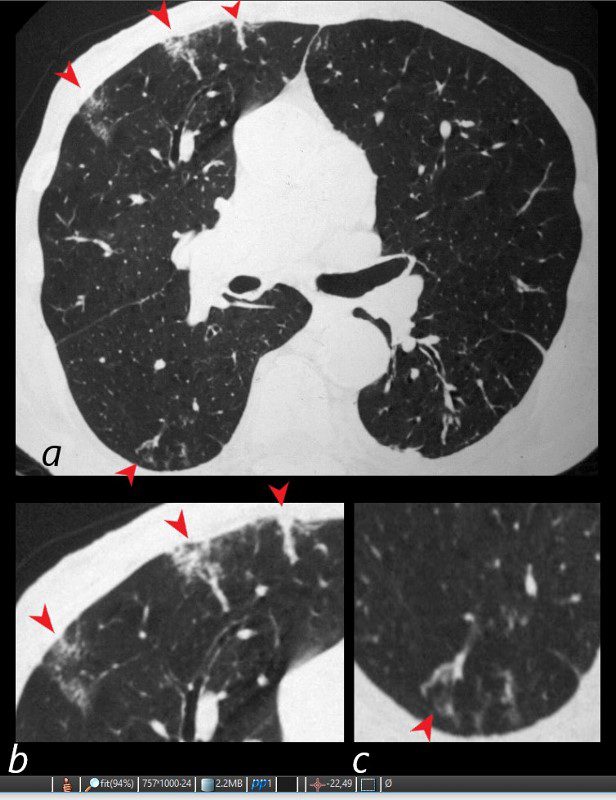
CT axial view (a) shows multiple peripheral wedge shaped ground glass densities subtended by distended feeding vessels (a,b,c, red arrowheads) reflecting areas of microinfarction due to vasculitis that affects both the arterioles and venules.
Priscilla Slanetz MPH MD
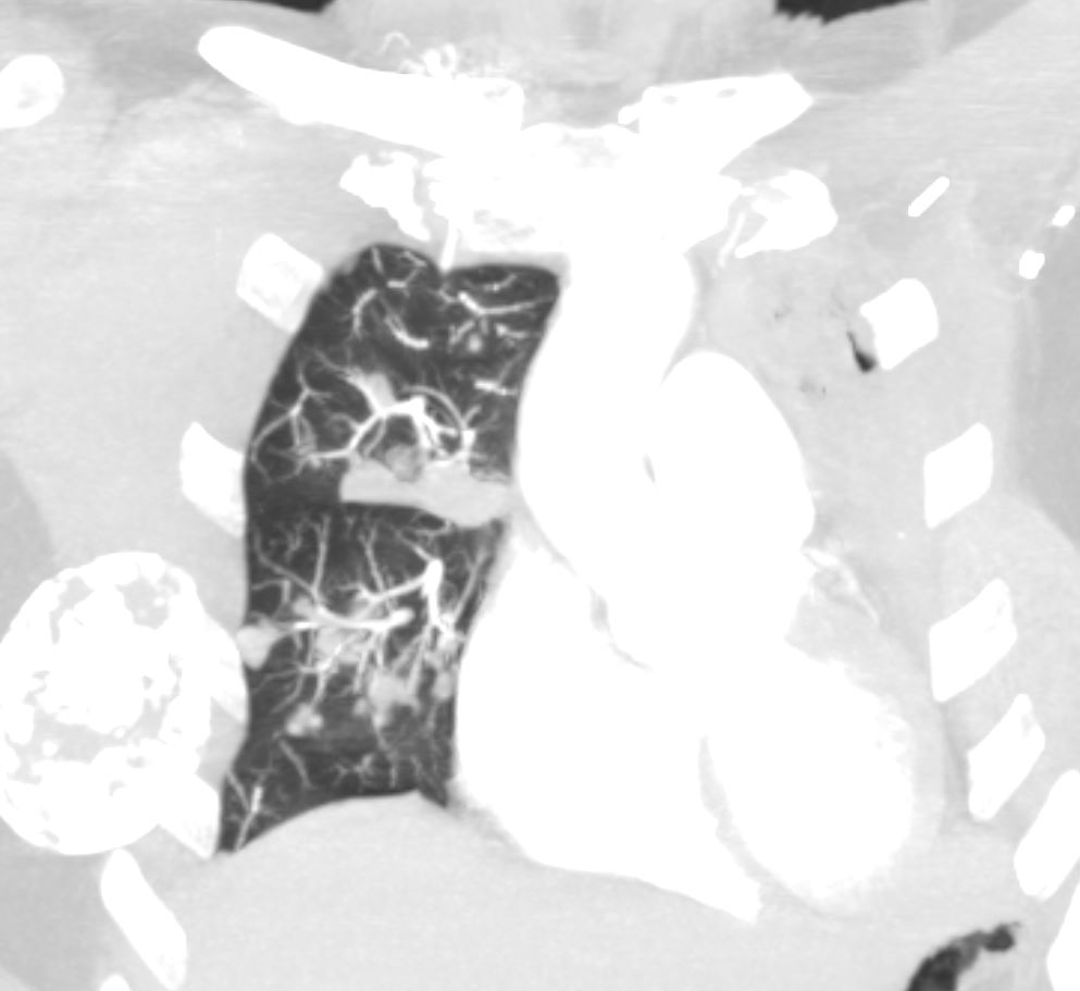
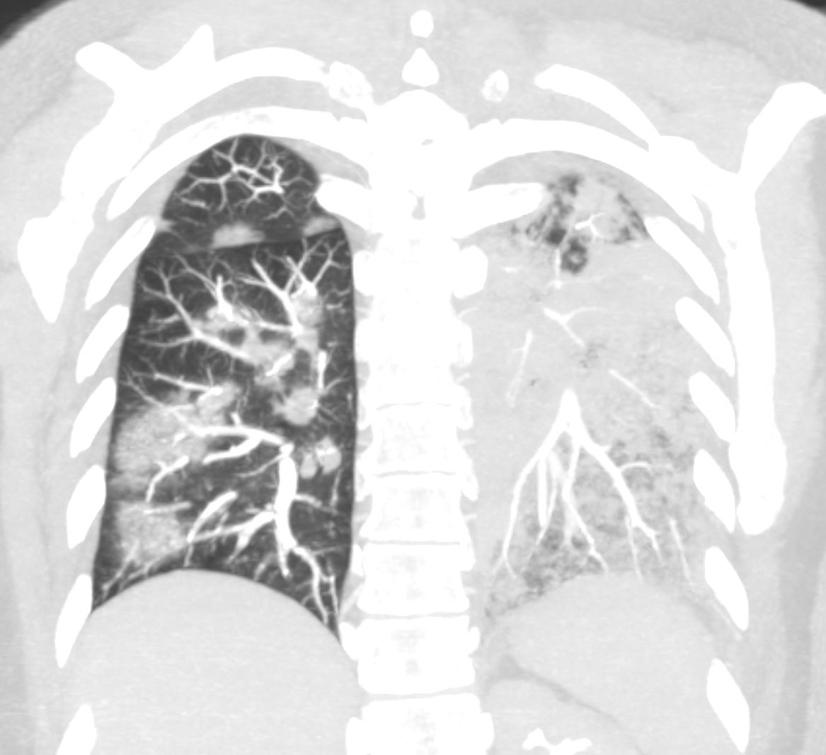
70F-year old female presents with hemoptysis and bilateral lower lobe pulmonary infiltrates CT scan shows a multiple nodules associated with blood vessels in the in the right lower lobe
Ashley Davidoff MD TheCommonVein.net Wegeners-cavitation-019
Ashley Davidoff MD TheCommonVein.net Wegeners-cavitation-021
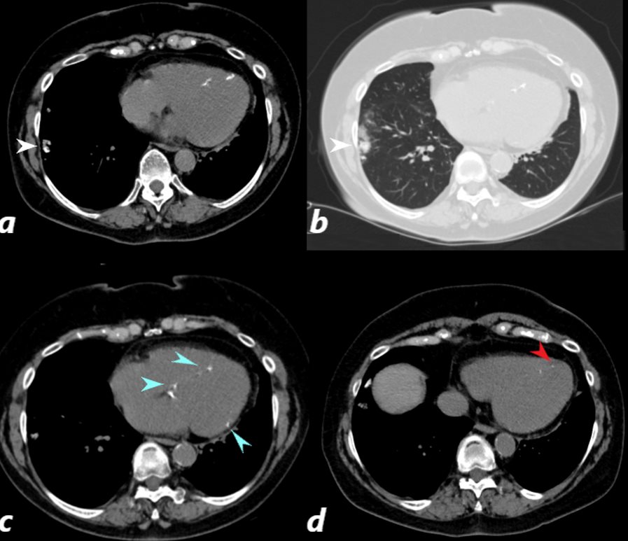
CT scan of a 67 year old female with anca vasculitis shows regions of dystrophic calcification in the lateral aspect of the right lower lobe (white arrow, a and b) )with focal nodular parenchymal consolidation, that likely reflects a site of prior small vessel infarct. Dystrophic calcification in the LV myocardium (blue arrows c) and a suggestion of fatty dysplasia in the left ventricular apex red arrow d) suggest changes from small vessel infarct. Ashley Davidoff MD TheCommonvein.net