
Source
Signs in Thoracic Imaging
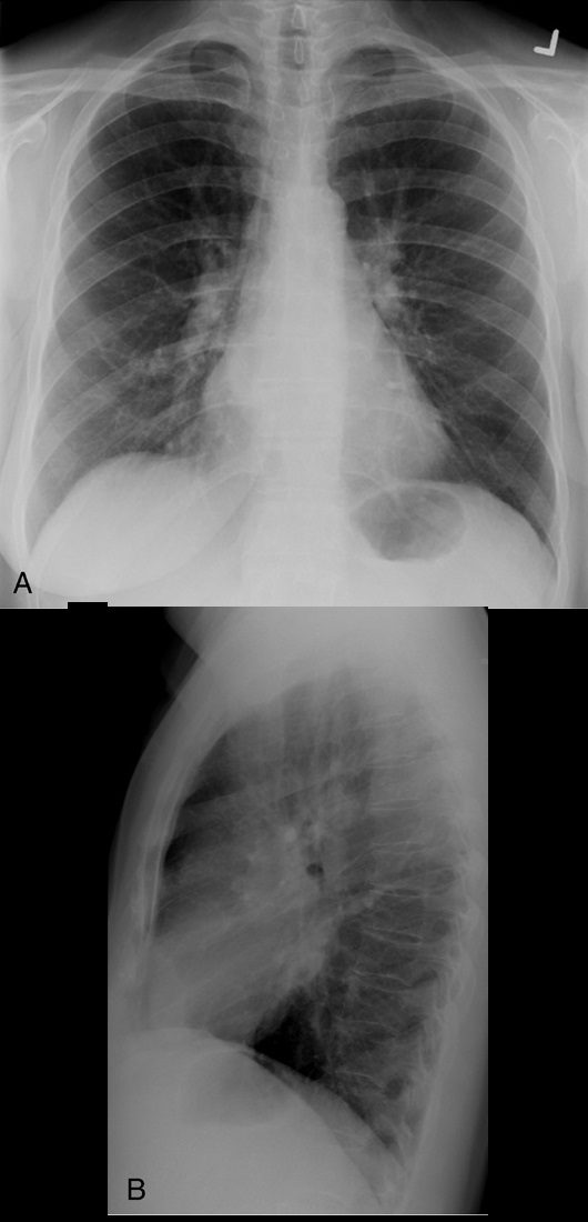
Journal of Thoracic Imaging Double density sign. Frontal radiograph (A) in a patient with known mitral stenosis shows a retrocardiac curvilinear density paralleling the right heart border, confirmed on the lateral view (B) to be due to left atrial enlargement.
Source
Signs in Thoracic Imaging
Journal of Thoracic Imaging 21(1):76-90, March 2006.21(1):76-90, March 2006.
On frontal chest radiographs, this sign presents as a curvilinear density projecting over the right retrocardiac region, signifying left atrial enlargement (Fig. 9).32 The curvilinear line represents the inferolateral margin of the left atrium.33 The double density sign may be observed in patients without cardiac disease; however, there is a semiquantitative measurement to estimate the left atrial diameter and better estimate whether it is a real finding.33 Higgins et al found that, on PA radiographs of adult patients, if the left atrial dimension is defined as the distance from the midpoint of the double density to the inferior wall of the left mainstem bronchus, a distance greater than 7 cm was consistent with a diagnosis of left atrial enlargement, confirmed on echocardiography.33 The measurement was found to be an unreliable sign in the evaluation of pediatric patients with a double density sign on PA radiographs.33