Nodules of Wegener’s

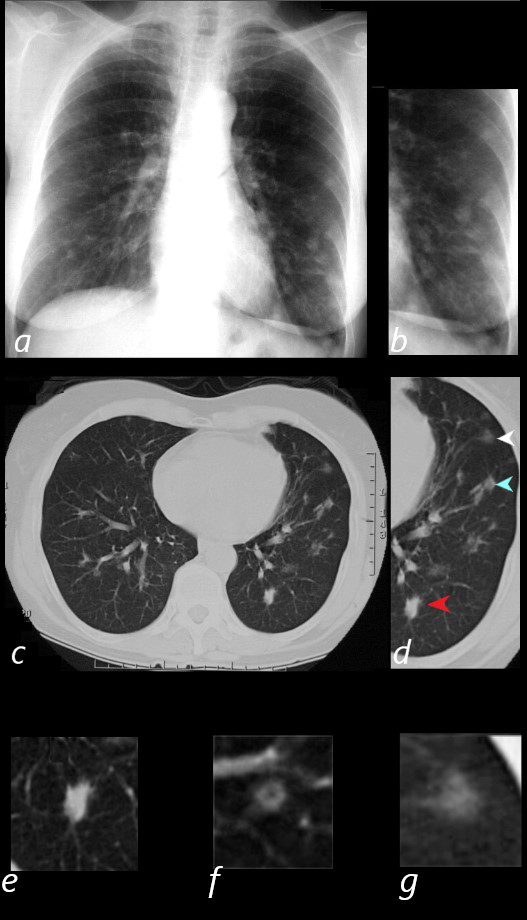
65 year old female presents with epistaxis and with nodular changes on CXR (a) magnified in b.
CT scan in axial projection (c) and magnified in d, reveals 3 types of nodules.
A spiculated solid nodule (red arrow head) is magnified in e, a bronchocentric nodule (teal arrowhead) is magnified in e. This may represent a cavitating nodule or hemorrhagic change around a bronchiole (cheerio sign) A ground glass nodule (white arrowhead) is magnified in g.
Ashley Davidoff MD

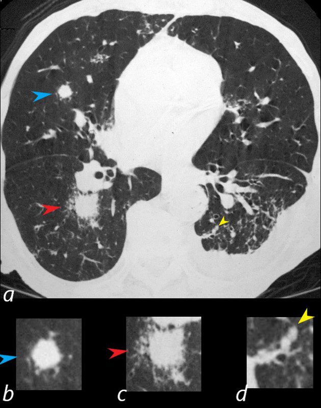
81-year-old male with weight loss, renal failure, and hemoptysis
CT axial view (a) shows a 1 cm solid nodule in the RUL surrounded by small vessels (a,b, blue arrowheads), a 1.8cm nodule in the superior segment of the right lower lobe with a halo sign indicating surrounding hemorrhage(a,c, red arrowheads), and a 3mm nodule in the superior segment of the left lower lobe (a,d, yellow arrowheads).
Priscilla Slanetz MPH MD

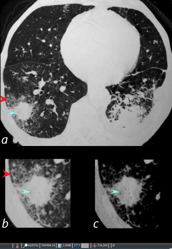
81-year-old male with weight loss, renal failure, and hemoptysis
CT axial view (a) shows a 2 cm solid nodule in the RUL surrounded by A HALO SIGN OF GROUND GLASS CHANGES AND RETICULAR CHANGES (a,b, red arrowheads), indicating surrounding hemorrhage, and subtle air bronchograms (a,b,c, teal arrowheads) best appreciated in c with narrowed windows.
Priscilla Slanetz MPH MD

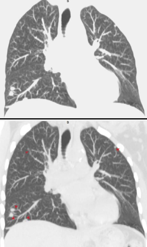
Coronal imaging shows multiple small nodules in the upper and lower lung field with noted subtending vessels
(feeding vessel sign)
Ashley Davidoff MD
-
Links and References
- TCV
- TCV
David NaegerUCSF
